结核与肺部疾病杂志 ›› 2021, Vol. 2 ›› Issue (2): 131-138.doi: 10.3969/j.issn.2096-8493.2021.02.008

• 论著 • 上一篇    下一篇

36例肺朗格汉斯细胞组织细胞增生症临床分析

黄伟华1, 王春燕2, 罗群1, 顾莹莹1, 谢佳星1, 夏亭亭3, 张清玲1, 张筱娴1()   

  1. 1广州医科大学附属第一医院 国家呼吸医学中心 国家呼吸系统疾病临床医学研究中心 呼吸疾病国家重点实验室 广州呼吸健康研究院呼吸与危重症医学科(黄伟华、罗群、顾莹莹、谢佳星、张清玲、张筱娴),广州 510120
    2广州医科大学附属第一医院血液科(王春燕),广州 510120
    3广州医科大学附属第一医院,放射科(夏亭亭),广州 510120
  • 收稿日期:2021-03-22 出版日期:2021-06-30 发布日期:2021-07-01
  • 通信作者: 张筱娴 E-mail:15002007401@163.com

Clinical analysis of 36 cases of pulmonary Langerhans cell histiocytosis

HUANG Wei-hua1, WANG Chun-yan2, LUO Qun1, GU Ying-ying1, XIE Jia-xing1, XIA Ting-ting3, ZHANG Qing-ling1, ZHANG Xiao-xian1()   

  1. 1Pulmonary and Critical Care Medicine, Guangzhou Institute of Respiratory Health, National Clinical Research Center for Respiratory Disease, National Center for Respiratory Medicine,State Key Laboratory of Respiratory Diseases, the First Affiliated Hospital of Guangzhou Medical University, Guangzhou 510120, China
    2Department of Hematology, the First Affiliated Hospital of Guangzhou Medical University,Guangzhou 510120,China
    3Department of Radiology, the First Affiliated Hospital of Guangzhou Medical University, Guangzhou 510120,China
  • Received:2021-03-22 Online:2021-06-30 Published:2021-07-01
  • Contact: ZHANG Xiao-xian E-mail:15002007401@163.com

摘要:

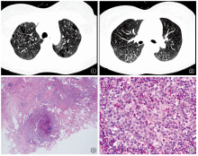
目的 探讨肺朗格汉斯细胞组织细胞增生症(pulmonary Langerhans cell histiocytosis,PLCH)的临床表现、影像学、病理及预后特征,以提高临床医生对该病的认识。方法 回顾性分析广州医科大学附属第一医院2009年9月至2019年8月收治住院的PLCH患者36例,对其临床资料进行分析。根据Muller评分法进行评分,对结节状病灶、囊状病变及肺间质性病变的HRCT评分进行比较。根据患者第1次行气管镜肺活检(transbronchial lung biopsy, TBLB)是否获得阳性结果将患者分为两组:阳性组(11例)和阴性组(8例);根据确诊PLCH的肺病理中浸润组织的嗜酸粒细胞的量分为两组:较多嗜酸粒细胞浸润组(18例)和较少嗜酸粒细胞浸润组(13例);根据患者的预后分为:存活组(32例)和死亡组(4例)。对两组间的临床特征进行比较。结果 36例PLCH患者发病中位年龄27.50(19.50,36.75)岁, 55.56%(20/36)的患者出现过自发性气胸,36例PLCH患者的HRCT总评分平均数为(6.71±2.84)分;结节状病灶、囊状病变及肺间质性病变的HRCT评分中位数分别为2.67(0.67,4.33)分、3.67(1.75,4.33)分和 0.00(0.00,1.92)分,且三者评分的差异具有统计学意义(χ2=18.000, P<0.001)。囊状病变HRCT评分与第1秒用力呼气容积占预计值的百分比 (the percentage of predicted value of forced expiratory volume in 1 second,FEV1%pred)及第1秒用力呼气容积/用力肺活量(forced expiratory volume in 1 second/forced vital capacity,FEV1/FVC)呈负相关(分别为:rs=-0.527,P=0.008; rs=-0.440,P=0.032)。19例患者进行TBLB,57.89%(11/19)第1次进行TBLB阳性组单次呼吸法肺一氧化碳弥散量占预计值的百分比(the percentage of predicted value of diffusion capacity for carbon monoxide of lung-single breath method,DLCO SB%pred)平均值为(75.19±11.91)%,明显优于阴性组[(55.43±17.10)%],差异有统计学意义(t=2.449,P=0.032);阳性组肺间质性病变HRCT评分均为0.00分,明显低于阴性组[4.50(0.00, 4.92)],差异有统计学意义(Z=-2.932,P=0.020)。确诊的PLCH肺病理组织中,有较多嗜酸粒细胞浸润组有61.11%(11/18)发现结节病灶,明显多于较少嗜酸粒细胞浸润组(15.38%)(Fisher精确概率法,P=0.025)。4例(11.11%,4/36)患者死亡,死亡组(100.00%)为多系统受累的PLCH,明显多于存活组(31.25%,10/32),差异有统计学意义(Fisher精确概率法,P=0.017)。结论 对于气胸为主要临床表现、囊状及结节状病变为主要影像学改变和/或伴有多系统受累的患者需考虑PLCH的可能;对于怀疑PLCH患者如弥散功能较好、HRCT中无间质性病变的表现,建议可尝试行TBLB确诊;多系统受累为预后不利因素。

关键词: 肺, 朗格汉斯细胞, 气胸, 嗜酸粒细胞增多, 病理学, 呼吸功能试验

Abstract:

Objective To improve the clinical understanding for pulmonary Langerhans cell histiocytosis (PLCH), including their clinical, radiological and pathological features and prognosis. Methods Retrospective analysis of 36 patients diagnosed with PLCH and hospitalized in The First Affiliated Hospital of Guangzhou Medical University between September 2009 and August 2019 was conducted. HRCT scores (using the Muller scoring method) of nodular lesions, cystic lesions and pulmonary interstitial lesions were compared. According to results of transbronchial lung biopsy (TBLB) at the first time,the patients were divided into two groups: positive TBLB result group (11 patients) and negative TBLB result group (8 patients). According to the level of eosinophilic infiltration in lung pathology,the patients were divided into two groups:higher eosinophilic infiltration group (18 patients) and lower eosinophilic infiltration group (13 patients). According to the prognosis, PLCH patients were divided into two groups: survival group (32 patients) and death group (4 patients).The clinical characteristics of those groups were compared. Results The median age of 36 patients with PLCH was 27.50 (19.50,36.75) years old. 55.56% (20/36) patients had spontaneous pneumothorax. The median HRCT total score of 36 patients was (6.71±2.84). The median HRCT scores of nodular lesions, cystic lesions and pulmonary interstitial lesions were 2.67 (0.67,4.33), 3.67 (1.75,4.33) and 0.00 (0.00,1.92), respectively, and the difference between them was statistically significant (χ2=18.000,P<0.001). HRCT score of cystic lesions was negatively correlated with the percentage of predicted value of forced expiratory volume in 1 second (FEV1%pred) and forced expiratory volume in 1 second/forced vital capacity (FEV1/FVC) (rs=-0.527,P=0.008;rs=-0.440,P=0.032, respectively). Nineteen patients underwent TBLB, and 57.89% (11/19) patients obtained positive pathological results of PLCH at the first time of TBLB. The average percentage of predicted value of diffusion capacity for carbon monoxide of lung-single breath method (DLCO SB%pred) among this positive TBLB result group was (75.19±11.91) %, significantly higher than that of the negative group ((55.43±17.10) %, t=2.449, P=0.032). The average HRCT score of pulmonary interstitial lesions in the positive group was 0.00, significantly lower than that in the negative group (4.50 (0.00, 4.92),P=0.020). In the lung pathological tissues of PLCH, 61.11% (11/18) of the patients with higher eosinophilic infiltration had nodular lesions, significantly higher than those with lower eosinophilic infiltration (15.38% (2/13),P=0.025). 4 (11.11%, 4/36) patients died and all the dead patients (100.00%) were diagnosed as multi-system PLCH, which was significantly higher than that in the survival group (31.25% (10/32),P=0.017). Conclusion For patients with pneumothorax as the main clinical manifestation, cystic and nodular lesions as the main radiological findings, with/without multi-system involvement, the possibility of PLCH should be considered. TBLB was recommended for suspected PLCH patients with better diffusion function and no interstitial lesions in HRCT. Multi-system involvement was the adverse factor of prognosis.

Key words: Lung, Pulmonary langerhans cell histiocytosis, Pneumothorax, Eosinophilia, Pathology, Respiratory Function Tests