结核与肺部疾病杂志 ›› 2020, Vol. 1 ›› Issue (3): 220-225.doi: 10.3969/j.issn.2096-8493.2020.03.004

• 论著 • 上一篇    下一篇

84例新型冠状病毒肺炎患者CT表现与免疫学指标的变化特点分析

刘伯飞*, 王芳 , 刘伯霞 , 马玉杰 , 冯涛 , 徐麟 , 赵桂霞 , 洪苑 , 刘广天 , 周攀 , 曹相原 ()   

  1. 750021 银川,宁夏回族自治区第四人民医院重症医学科(刘伯飞、王芳、赵桂霞、洪苑),呼吸一科(徐麟),防治科(刘广天),放射科(周攀);宁夏医科大学基础医学院(刘伯霞);宁夏医科大学总医院重症医学科(马玉杰、曹相原);宁夏回族自治区第三人民医院重症医学科(冯涛)
  • 收稿日期:2020-10-09 出版日期:2020-12-30 发布日期:2021-01-05
  • 通信作者: 曹相原 E-mail:c_xyuan@hotmail.com
  • 基金资助:
    宁夏回族自治区新型冠状病毒感染的肺炎疫情科技项目(2020GZBD0003);宁夏回族自治区重点研发基金项目(2020BEG03012)

Analysis of characteristics of chest CT imaging and immune indexes in 84 COVID-19 patients

LIU Bo-fei*, WANG Fang , LIU Bo-xia , MA Yu-jie , FENG Tao , XU Lin , ZHAO Gui-xia , HONG Yuan , LIU Guang-tian , ZHOU Pan , CAO Xiang-yuan ()   

  1. *Department of Critical Medicine,the Fourth People’s Hospital of Ningxia Hui Autonomous Region, Yinchuan 750001, China
  • Received:2020-10-09 Online:2020-12-30 Published:2021-01-05
  • Contact: CAO Xiang-yuan E-mail:c_xyuan@hotmail.com

摘要:

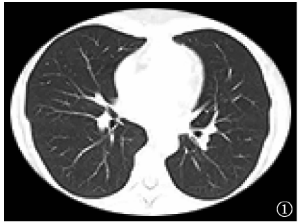
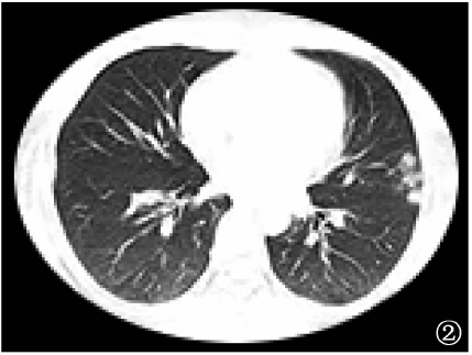
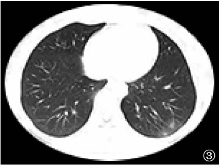
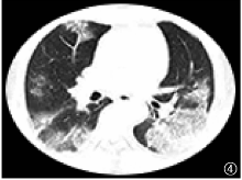
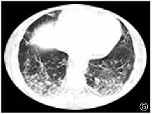
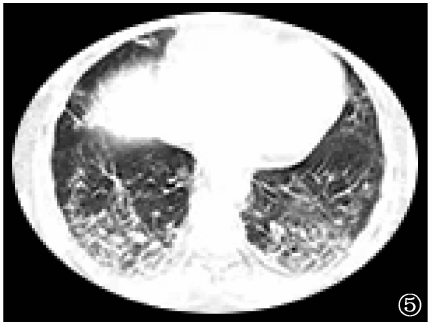
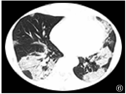
目的 分析新型冠状病毒肺炎( coronavirus disease 2019,COVID-19 ) 患者肺部影像学和免疫学指标的变化特点,为早期判定疾病进展提供参考。方法 回顾性收集宁夏回族自治区( 简称“宁夏” ) 2020年1月24日至3月7日收治的COVID-19确诊患者84例,其中75例为确诊患者[有明确流行病学史、发热和( 或 ) 肺部典型CT扫描表现,以及核酸检测阳性者],9例临床诊断患者( 有明确流行病学史或密切接触者、存在典型肺部影像学改变,但两次核酸检测阴性者 ) 。根据动态监测的胸部CT表现的变化和实验室免疫学指标检查结果,分析COVID-19患者入院72h时的胸部CT扫描表现的变化与患者临床分型及免疫状态之间的关系。结果 患者入院24h内,轻型、普通型和重/危重型分别为14、59和11例,其中普通型胸部CT表现的双肺间质性病灶[1.7%( 1/59 ) ]和双肺弥漫性病灶[5.1%( 3/59 ) ]明显低于重/危重型患者[分别为18.2%( 2/11 ) 和27.3%( 3/11 ) ]( χ2=6.144,P=0.013;χ2=5.824,P=0.016 ) 。入院72h后,有9例( 10.7% ) 患者临床分型发生进展,轻型、普通型和重/危重型患者分别为10、58、16例,其中普通型胸部CT表现的少量/偶发片状病灶[20.7%( 12/58 ) ]和双肺多发病灶[62.1%( 36/58 ) ]明显高于重/危重型患者[分别为0.0%( 0/16 ) 和18.8%( 3/16 ) ]( χ2=3.951,P=0.047;χ2=9.441,P=0.002 ) ;而肺间质病灶[0.0%( 0/58 ) ]和双肺弥漫性病灶[8.6%( 5/58 ) ]明显低于重/危重型患者[31.2%( 5/16 ) 和50.0%( 8/16 ) ]( χ2=19.438,P<0.001;χ2=14.828,P<0.001 ) 。对CT表现为病灶恶化的43例和无恶化的41例患者进行相关免疫指标检测,结果显示:恶化组患者WBC计数的中位数( 四分位数 ) [M( Q1,Q3 ) ]为4.460( 3.560,4.900 ) ×109/L、淋巴细胞总数为1.290( 0.900,1.520 ) ×109/L、CD3+为496.000( 304.000,802.000 ) /μl、CD3+CD4+为325.000( 183.000,480.000 ) /μl、CD3+CD8+为186.000( 99.000,330.000 ) /μl、CD3+CD4+CD8+为2.000( 1.000,5.000 ) /μl、CD45+为998.000( 500.000,1198.000 ) /μl,均明显低于无恶化组的M( Q1,Q3 ) [分别为5.130( 4.225,7.050 ) ]×109/L、1.600( 1.295,2.090 ) ×109/L、1001.000( 766.500,1230.000 ) /μl、590.000( 468.500,765.000 ) /μl、380.000( 227.500,535.000 ) /μl、10.000( 5.000,18.000 ) /μl、1530.000( 1064.000,1885.000 ) /μl]( U值分别为542.500,503.500,348.000,348.000,457.000,261.000,359.000,P值分别为0.002,<0.001,<0.001,<0.001,<0.001,<0.001,<0.001 ) 。而恶化组补体C3的M( Q1,Q3 ) 为1.200( 1.000,1.330 ) g/L,明显高于无恶化组的1.060( 0.960,1.225 ) g/L( U=1118.500,P=0.034 ) 。结论 COVID-19患者入院72h后肺部病灶是否恶化可能与免疫状态改变相关,监测CT扫描肺部病灶的变化及免疫学指标的改变对了解疾病进展、早期开展预防性诊治具有指导意义。

关键词: 新型冠状病毒肺炎, 体层摄影术,X线计算机, 监测,免疫学, 疾病恶化, 疾病特征, 对比研究, 数据说明,统计

Abstract:

Objective To anlyze the characteristics of chest CT imaging and immune indexes in patients with COVID-19. Methods A total of 84 COVID-19 patients in Ningxia between January 24, 2020 and March 7, 2020 were retrospectively studied, including 75 confirmed cases (difinite epidemiological history, fever and/or typical chest CT images, and positive in nucleic acid test) and 9 clinically diagnosed cases (difinite epidemiological history or close contact, typical chest CT imaging, but negative in two nucleic acid tests). According to the dynamic CT imaging and results of immune indexes, the relationship between the changes of chest CT 72 hours after admission and the clinical classification and immune status of COVID-19 patients were analyzed. Results Within 24 hours of admission, the 84 patients were divided into mild (n=14), ordinary (n=59), and severe/critical types (n=11). By CT scan, the proportions of interstitial lesions in bilateral lung and diffuse lesions in bilateral lung in ordinary patients were significantly lower than those in severe/critical patients (1.7% (1/59) vs. 18.2% (2/11), χ2=6.144, P=0.013; 5.1% (3/59) vs. 27.3% (3/11), χ 2=5.824, P=0.016). The classification of 9 patients was progressing 72 hours after admission, therefore, numbers of mild, ordinary, and severe/critical types were changed to 10, 58 and 16, respectively. By CT scan, compared with those in patients of severe/critical type, few/occasional patchy lesions lesions and multiple lesions in bilateral lung in patients of ordinary type were significantly higher (20.7% (12/58) vs.0.0% (0/16), χ 2=3.951, P=0.047; 62.1% (36/58) vs. 18.8% (3/16), χ 2=9.441, P=0.002); while interstitial lesionsn and diffuse lesions in bilateral lung in patients of ordinary type were significantly lower (0.0% (0/58) vs. 31.2% (5/16), χ 2=19.438, P<0.001; 8.6% (5/58) vs. 50.0% (8/16), χ 2=14.828, P<0.001).The relevant immune tests were performed on 43 patients with deterioration (deterioration group) in CT and 41 patients without deterioration (non-deterioration group). It was found that, the WBC count (M(Q1,Q3)), T lymphocyte, CD3 +, CD3+CD4+, CD3+CD8+, CD3+CD4+CD8+ and CD45+ cells in deterioration group were siginificantly lower than those in non-deterioration group (4.460 (3.560, 4.900)×109/L vs. 5.130 (4.225, 7.050)×10 9/L, 1.290 (0.900, 1.520)×109/L vs. 1.600 (1.295,2.090)×10 9/L, 496.000 (304.000, 802.000)/μl vs. 1001.000 (766.500, 1230.000)/μl, 325.000 (183.000,480.000)/μl vs. 590.000 (468.500, 765.000)/μl, 186.000 (99.000, 330.000)/μl vs. 380.000.(227.500, 535.000)/μl, 2.000 (1.000, 5.000)/μl vs. 10.000 (5.000, 18.000)/μl, 998.000 (500.000, 1198.000)/μl vs. 1530.000 (1064.000,1885.000)/μl; U=542.500, 503.500, 348.000, 348.000, 457.000, 261.000, and 359.000, respectively; PWBC=0.002, and all of others P<0.001). Complement C3 in deterioration group was siginificantly higher than that in non-deterioration group (1.200 (1.000, 1.330) g/L vs. 1.060 (0.960, 1.225) g/L; U=118.500, P=0.034). Conclusion The deterioration of pulmonary lesions in patients with COVID-19 within 72 hours after admission may be related to the changes of immune status. Monitoring the changes of chest CT and immune indexes is of guiding significance for diagnosis and treatment.

Key words: Coronavirus disease 2019 (COVID-19), Tomography,X-ray computer, Monitoring,immunologic, Disease progression, Disease attributes, Comparative study, Data interpretation,statistical