结核病与肺部健康杂志 ›› 2020, Vol. 9 ›› Issue (1): 25-31.doi: 10.3969/j.issn.2095-3755.2020.01.007

• 论著 • 上一篇    下一篇

新型冠状病毒肺炎患者不同发病时期肺周病变超声成像表现的初步探讨

黄毅, 王思翰, 郑楚云, 刘月, 张耀辉()   

  1. 710010 西安市胸科医院超声科(黄毅、王思翰、郑楚云、刘月),医务科统计室(张耀辉)
  • 收稿日期:2020-03-13 出版日期:2020-03-30 发布日期:2020-04-17
  • 通信作者: 张耀辉 E-mail:328162296@qq.com

Ultrasonographic features of peripheral lung lesions at different stages in patients with novel coronavirus pneumonia

HUANG Yi, WANG Si-han, ZHENG Chu-yun, LIU Yue, ZHANG Yao-hui()   

  1. Department of Ultrasound, Xi’an Chest Hospital, Xi’an 710100, China
  • Received:2020-03-13 Online:2020-03-30 Published:2020-04-17
  • Contact: ZHANG Yao-hui E-mail:328162296@qq.com

摘要:

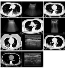
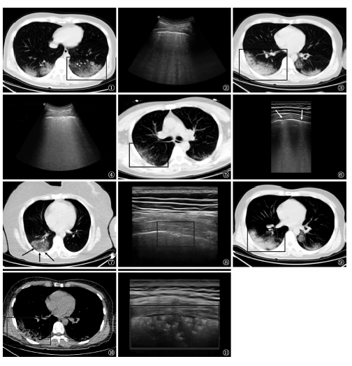
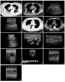
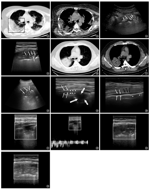
目的 探讨新型冠状病毒肺炎[WHO命名为“Corona virus disease 2019(COVID-19)”]患者不同发病时期肺周病变的超声成像表现,为临床诊断及疗效评估提供参考。方法 分析2020年1—2月份西安市胸科医院收治经临床确诊的COVID-19患者共15例。其中,早期患者3例,进展期患者10例,重症期患者2例,均行常规二维超声及彩色多普勒超声检查,观察病灶超声成像特征。结果(1)主要超声表现为胸膜线欠光滑或中断,产生实变影和可见到B线,发生率为100.00%(15/15)。其次为实变病灶内支气管充气征,发生率为66.67%(10/15),胸膜增厚约为53.33%(8/15)。(2)不同发病时期患者肺周病变的超声成像有不同特征。早期患者可见肺周胸膜线毛糙欠光滑;进展期患者可见患者胸膜线毛糙欠光滑,有部分中断,同时可见数条B线,且B线位置固定,部分患者胸膜下可见小斑片状肺实变影;重症期患者除普通型患者表现特征外,可见胸膜线中断消失,胸膜下大片不规则实变影及大量融合B线,在实变区域内可见支气管充气征,同时病灶相邻胸膜下可有局限性增厚,病灶实变区域彩色多普勒血流成像可显示乏血流信号。结论 COVID-19患者的超声表现在病情不同进展时期具有一定特征性,可为临床诊断及疗效评估提供依据。

关键词: 新型冠状病毒肺炎, 超声检查, 诊断显像, 疾病特征

Abstract:

Objective To investigate the ultrasonographic features of peripulmonary lesion of novel coronavirus pneumonia (COVID-19) patients in different period of onset, and to provide reference for clinical diagnosis and efficacy evaluation. Methods Retrospective analysis was conducted in the 15 patients with COVID-19 diagnosed clinically in our hospital from January to February 2020. Among them, 3 patients were in early stage, 10 patients were in advanced stage and 2 patients were in severe stage. All patients were examined by two-dimensional ultrasound and color doppler ultrasound to observe the image characteristics of lesions under ultrasound imaging. Results (1) The main manifestations of ultrasound were that the pleura line was not smooth or interrupted, resulting in solid shadows and visible “B” line. The incidence of the above three ultrasonic imaging changes was 100.00% (15/15), bronchiectasis in solid lesions was 66.67% (10/15) and pleural thickening was 53.33% (8/15). (2) There were different characteristics in different stages of the disease. It was occasionally seen that the peripulmonary pleura line was rough in the early stage of the disease. In the advanced stage, the pleura line of the patients was rough and partial interruption, at the same time several B lines could be seen and the position of B line was fixed, in some patients subpleural patchy pulmonary consolidation could be seen. In the severe stage, beside the characteristics of common patients, interruption and disappearance of pleura line, large irregular consolidation shadow under pleura and a large number of fusion B line were observed, as well as bronchiectasis in the consolidation area. At the same time, the adjacent pleura had local thickening. Color doppler flow imaging (CDFI) in the focal consolidation area showed lack of blood flow signals. Conclusion The ultrasound images of patients with COVID-19 have certain characteristics imaging manifestations at different stages of disease progression, which can provide basis for clinical diagnosis and efficacy evaluation.

Key words: Coronavirus disease 2019(COVID-19), Ultrasonography, Diagnostic imaging, Disease attributes