结核病与肺部健康杂志 ›› 2020, Vol. 9 ›› Issue (1): 58-63.doi: 10.3969/j.issn.2095-3755.2020.01.013

• 论著 • 上一篇    下一篇

儿童耐药肺结核的CT特征分析

宋敏, 方伟军(), 韩远远, 冯惠勇   

  1. 510095 广州市胸科医院放射科
  • 收稿日期:2019-10-31 出版日期:2020-03-30 发布日期:2020-04-17
  • 通信作者: 方伟军 E-mail:13533336916@163.com
  • 基金资助:
    广州市高水平临床重点专科和培育专科建设项目(穗卫函[2019]1555号);广东省转化医学创新平台培育建设项目B类(惠卫函[2018]1254号)

Analysis of CT features of drug-resistant tuberculosis in children

SONG Min, FANG Wei-jun(), HAN Yuan-yuan, FENG Hui-yong   

  1. Department of Radiology, Guangzhou Chest Hospital, Guangzhou 510095, China
  • Received:2019-10-31 Online:2020-03-30 Published:2020-04-17
  • Contact: FANG Wei-jun E-mail:13533336916@163.com

摘要:

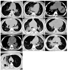
目的 探讨儿童耐药肺结核患者的CT特征。方法 收集广州市胸科医院2012年1月至2018年12月经临床及实验室确诊的儿童耐药肺结核29例为耐药肺结核组(DR组);采用1∶3病例对照研究,选取同期符合纳入标准的对抗结核药物敏感的结核病患儿87例,归为敏感组(DS组)。匹配原则为同性别、年龄±2岁。两组患儿按照年龄将0~岁患儿分为耐药1组(DR1组)12例、敏感1组(DS1组)36例,将 5~14岁患儿分为耐药2组(DR2组)17例、敏感2组(DS2组)51例,对比分析不同年龄段对抗结核药物耐药患儿与敏感患儿的CT特征,总结耐药结核病儿童的CT表现特征。结果 DR1组与DS1组、DR2组与DS2组的肺内病灶累及双肺各叶的发生率分别为66.67%(8/12)和30.56%(11/36)、52.94%(9/17)和23.53%(12/51),差异均有统计学意义(χ2值分别为4.907、5.167,P值分别为0.027、0.023)。DR1组及DS1组肺内多发结节状病灶的发生率分别为83.33%(10/12)、44.44%(16/36),肺实变发生率分别为50.00%(6/12)、86.11%(31/36),出现胸腔积液者分别为8.33%(1/12)、38.89%(14/36),支气管狭窄发生率分别为16.67%(2/12)、52.78%(19/36),差异均有统计学意义(χ2值分别为5.483、6.644、3.911、4.769,P值分别为0.019、0.010、0.048、0.029)。DR2组及DS2组胸腔积液的发生率分别为11.76%(2/17)、43.14%(22/51),支气管狭窄的发生率分别为47.06%(8/17)、21.57%(11/51),两组比较差异均有统计学意义(χ2值分别为5.495、4.115,P值分别为0.019、0.043)。结论 DR组的CT特征是肺内病变范围较DS组广泛;0~岁年龄组的耐药肺结核患儿的肺内以多发结节状病灶常见;5~14岁年龄组的耐药肺结核患儿的支气管狭窄发生率较高。

关键词: 儿童, 结核,肺, 结核,抗多种药物性, 体层摄影术,螺旋计算机, 年龄组, 疾病特征, 数据说明,统计

Abstract:

Objective To investigate the CT imaging features of drug-resistant tuberculosis (DR-TB) in children. Methods A total of 29 children with DR-TB in Guangzhou Chest Hospital diagnosed by clinical and laboratory from January 2012 to December 2018 were retrospectively collected as DR-TB group (Group DR). Meanwhile, based on 1:3 case-control study, 87 children in the same period with drug-sensitive tuberculosis (DS-TB) who met the inclusion criteria were selected as drug-sensitive group (Group DS). The principle of comparative matching was the same sex and age ±2 years old. In these two groups, the children aged 0- years old were classified into Drug-resistant Group 1 (Group DR1, 12 cases), and Drug-sensitive Group 1 (Group DS1, 36 cases); the children aged 5-14 years old were classified into Drug-resistant Group 2 (Group DR2, 17 cases), and Drug-sensitive Group 2 (Group DS2, 51 cases). The CT features of drug-resistant children and drug-sensitive children in different age groups were compared and analyzed, and the CT features of DR-TB children were summarized. Results The incidence of lesions involving lobes of both lungs in Group DR1 and Group DS1, Group DR2 and Group DS2 was 66.67% (8/12) and 30.56% (11/36), 52.94% (9/17) and 23.53% (12/51), respectively. There were significantly differences between the two groups (χ2 values: 4.907, and 5.167, respectively; P values: 0.027 and 0.023, respectively). The incidence of multiple nodular lesions in Group DR1 and Group DS1 was 83.33% (10/12) and 44.44% (16/36), respectively, and that of consolidation was 50.00% (6/12) and 86.11% (31/36), that of pleural effusion was 8.33% (1/12) and 38.89% (14/36), that of bronchostenosis was 16.67% (2/12) and 52.78% (19/36), respectively. There were significantly differences in these aspects between the two groups (χ2 values: 5.483, 6.644, 3.911, and 4.769, respectively; P values: 0.019, 0.010, 0.048, and 0.029, respectively). The incidence of pleural effusion in Group DR2 and Group DS2 was 11.76% (2/17) and 43.14% (22/51), respectively, and that of bronchostenosis was 47.06% (8/17) and 21.57% (11/51), respectively. There were significantly differences between the two groups (χ2 values: 5.495, and 4.115, respectively; P values: 0.019 and 0.043, respectively). Conclusion The CT features of children with DR-TB are as follows: compared with children with DS-TB in the same age group, lung lesions are more widely distributed in children with DR-TB; multiple nodular lesions are common in DR-TB children aged 0- years old, and there is high incidence of bronchostenosis in DR-TB children aged 5-14 years old.

Key words: Child, Tuberculosis, pulmonary, Tuberculosis, multidrug-resistant, Tomography, spiral computed, Age groups, Disease attributes, Data interpretation, statistical