结核与肺部疾病杂志 ›› 2023, Vol. 4 ›› Issue (6): 486-492.doi: 10.19983/j.issn.2096-8493.20230106

• 论著 • 上一篇    下一篇

肺结核合并肺栓塞患者影像学特征分析

刘蕾1, 崔灿灿1, 李明武2(), 万荣2, 刘颖楠2   

  1. 1大理大学公共卫生学院,大理 671000
    2昆明市第三人民医院结核二科,昆明 650041
  • 收稿日期:2023-09-02 出版日期:2023-12-20 发布日期:2023-12-18
  • 通信作者: 李明武,Email:ynkmlmw@sina.com
  • 基金资助:
    云南省教育厅科学研究基金(2022J0721)

CT image analysis of patients with pulmonary tuberculosis complicated with pulmonary embolism

Liu Lei1, Cui Cancan1, Li Mingwu2(), Wan Rong2, Liu Yingnan2   

  1. 1School of Public Health, Dali University, Yunnan Province, Dali 671000, China
    2The Second Department of Tuberculosis, Kunming Third People’s Hospital, Yunnan Province, Kunming 650041, China
  • Received:2023-09-02 Online:2023-12-20 Published:2023-12-18
  • Contact: Li Mingwu, Email: ynkmlmw@sina.com
  • Supported by:
    Scientific Research Fund Project of Education Department of Yunnan Province(2022J0721)

摘要:

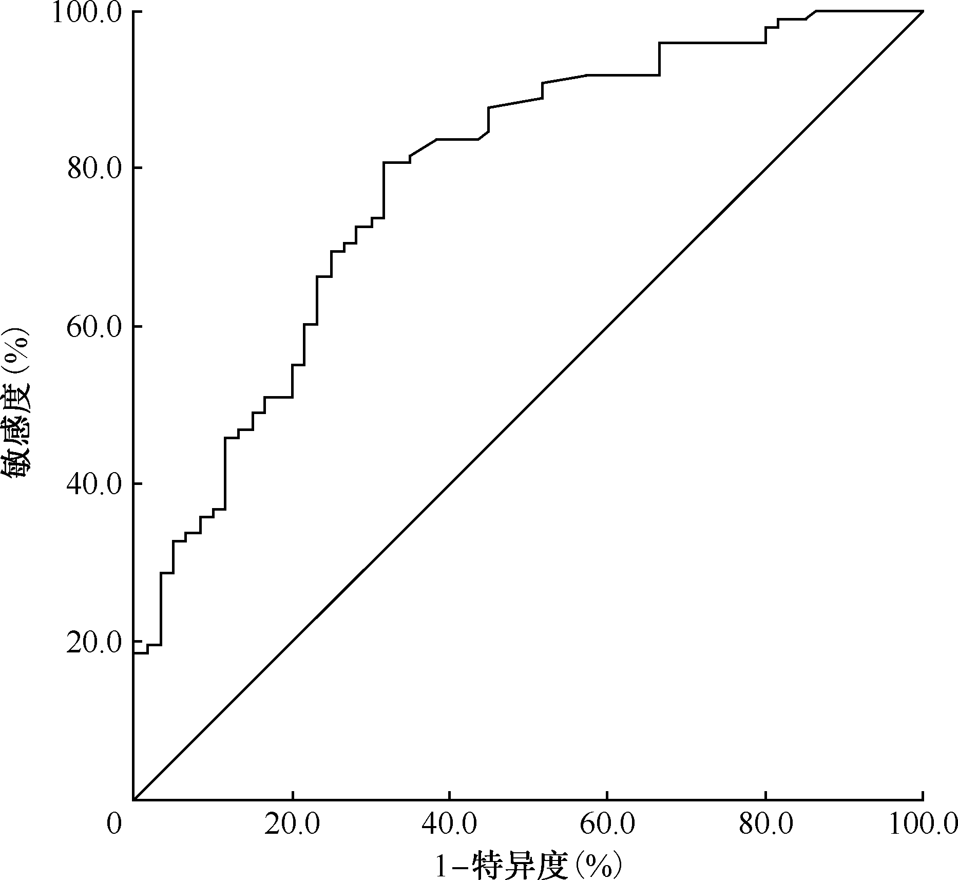
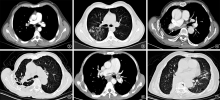
目的: 分析肺结核合并肺栓塞患者胸部CT影像学表现,提高临床医生对该病影像学的认识。方法: 回顾性选择2017年1月至2022年12月昆明市第三人民医院确诊为肺结核合并肺栓塞的101例患者为观察组,从同期确诊为单纯肺结核的39495例患者中,通过Excel软件生成随机数字,抽取101例患者作为对照组,收集两组患者的一般资料及影像学资料,对病灶分布和形态、病变类型和部位进行总结分析。结果: 观察组中位年龄[65.00(51.00,71.50)岁]高于对照组[39.00(27.00,55.00)岁],复治患者(58例,57.4%)占比高于对照组(33例,32.7%),差异均有统计学意义(Z=―7.562,P<0.001;χ2=12.499,P<0.001);观察组栓子分布于右下叶者最多,为43例(42.6%,43/101);观察组病变分布于右上叶、右中叶、右下叶、左上叶、左下叶者分别为53例(52.5%,53/101)、26例(25.7%,26/101)、49例(48.5%,49/101)、50例(49.5%,50/101)和43例(42.6%,43/101),与对照组[50例(49.5%,50/101)、19例(18.8%,19/101)、39例(38.6%,39/101)、43例(42.6%,43/101)和37例(36.6%,37/101)]相比,差异均无统计学意义(χ2=0.339,P=0.560;χ2=1.616,P=0.204;χ2=2.702,P=0.100;χ2=1.410,P=0.235;χ2=1.020,P=0.312);观察组病变范围累积三叶及以上,影像学表现为斑片影、实变影,发生毁损肺者分别为70例(69.3%,70/101)、56例(55.4%,56/101)、12例(11.9%,12/101)和37例(36.6%,37/101),均多于对照组[36例(35.6%,36/101)、39例(38.6%,39/101)、2例(2.0%,2/101)和4例(4.0%,4/101)],差异均有统计学意义(χ2=31.439,P<0.001;χ2=6.012,P=0.014;χ2=8.176,P=0.004;χ2=33.325,P<0.001);观察组伴有肺气肿、心包积液、双侧胸腔积液、右心增大、肺不张、肺动脉增粗者分别为29例(28.7%,29/101)、30例(29.7%,30/101)、43例(42.6%,43/101)、37例(36.6%,37/101)、17例(16.8%,17/101)和33例(32.7%,33/101),均多于对照组[5例(5.0%,5/101)、4例(4.0%,4/101)、8例(7.9%,8/101)、4例(4.0%,4/101)、6例(5.9%,6/101)和3例(3.0%,3/101)],差异均有统计学意义(χ2=20.688,P<0.001;χ2=24.243,P<0.001;χ2=33.635,P<0.001;χ2=49.155,P<0.001;χ2=6.488,P=0.011;χ2=33.223,P<0.001)。受试者工作特征曲线显示,当患者D-二聚体>1.290μg/ml时,肺结核患者发生肺栓塞的可能性较大。结论: 在年龄较高的复治肺结核患者中,出现斑片影、实变影、毁损肺,同时伴有肺气肿、心包积液、双侧胸腔积液、右心增大、肺不张、肺动脉增粗和D-二聚体>1.290μg/ml时,要警惕肺栓塞的发生,应结合患者的临床症状及其他实验室检查结果,对疾病进行早诊断和早治疗。

关键词: 结核, 肺, 肺栓塞, 体层摄影术, X线计算机, 对比研究

Abstract:

Objective: To analyze the chest CT imaging manifestations of patients with pulmonary tuberculosis complicated with pulmonary embolism, and to improve clinicians’ understanding of the imaging of this disease. Methods: A total of 101 patients diagnosed with pulmonary tuberculosis combined with pulmonary embolism in the Kunming Third People’s Hospital from January 2017 to December 2022 were retrospectively selected as the observation group (all patients admitted during this period), and 101 patients diagnosed with simple pulmonary tuberculosis were selected as the control group from 39495 patients by random numbers generated by Excel. The general data and chest CT imaging data were collected, and the distribution, morphology, lesion type and location were summarized and analyzed. Results: The median age of the observation group (65.00 (51.00, 71.50) years) was higher than that of the control group (39.00 (27.00, 55.00) years) (Z=―7.562, P<0.001). Proportion of retreatment patients (57.4%, 58/101) in the observation group was statistically higher than that (32.7%, 33/101) in the control group (χ2=12.499, P<0.001). In the observation group, 43 cases (42.6%, 43/101) had emboli located in the lower right lung lobe. The distribution of lesions in the observation group were 53 cases (52.5%, 53/101) in the right upper lobe, 26 cases (25.7%, 26/101) in the right middle lobe, 49 cases (48.5%, 49/101) in the right lower lobe, 50 cases (49.5%, 50/101) in the left upper lobe, 43 cases (42.6%, 43/101) in the left lower lobe, while for the control group were 50 cases (49.5%, 50/101), 19 cases (18.8%, 19/101), 39 cases (38.6%, 39/101), 43 cases (42.6%, 43/101), 37 cases (36.6%, 37/101), the differences were not statistically significant (χ2=0.339, P=0.560; χ2=1.616, P=0.204; χ2=2.702, P=0.100; χ2=1.410, P=0.235; χ2=1.020, P=0.312); In the observation group, 70 cases (69.3%, 70/101) had lesions accumulated in three or more than three lobes, 56 cases (55.4%, 56/101) showed patchy shadow, 12 cases (11.9%, 12/101) showed consolidation shadow, 37 cases (36.6%, 37/101) showed destroyed lungs, they were all higher than that in the control group (36 cases (35.6%, 36/101), 39 cases (38.6%, 39/101), 2 cases (2.0%, 2/101) and 4 cases (4.0%, 4/101)), and the differences were all statistically significant (χ2=31.439, P<0.001; χ2=6.012, P=0.014; χ2=8.176, P=0.004; χ2=33.325, P<0.001); In the observation group, there were 29 cases (28.7%, 29/101) complicated with emphysema, 30 cases (29.7%, 30/101) with pericardial effusion, 43 cases (42.6%, 43/101) with bilateral pleural effusion, 37 cases (36.6%, 37/101) with right heart enlargement, 17 cases (16.8%, 17/101) with atelectasis, and 33 cases (32.7%, 33/101) with pulmonary artery thickness, which were all statistically higher than that of the control group (5 cases (5.0%, 5/101), 4 cases (4.0%, 4/101), 8 cases (7.9%, 8/101), 4 cases (4.0%, 4/101), 6 cases (5.9%, 6/101), 3 cases (3.0%, 3/101); χ2=20.688, P<0.001; χ2=24.243, P<0.001; χ2=33.635, P<0.001; χ2=49.155, P<0.001; χ2=6.488, P=0.011; χ2=33.223, P<0.001). The working characteristic curves showed that when D-dimer >1.290 μg/ml, pulmonary tuberculosis patients were more likely to develop pulmonary embolism. Conclusion: In older patients with retreated pulmonary tuberculosis whose CT show patchy shadow, consolidation shadow or damaged lungs, accompanied by emphysema, pericardial effusion, bilateral pleural effusion, right heart enlargement, atelectasis, pulmonary artery thickening and D-dimer >1.290 μg/ml, it is necessary to be alert to the occurrence of pulmonary embolism. Doctors should consider clinical symptoms and other laboratory examinations of patients to make early diagnosis and treatment.

Key words: Tuberculosis, pulmonary, Pulmonary embolism, Tomography, X-ray computed, Comparative study

中图分类号: