结核与肺部疾病杂志 ›› 2022, Vol. 3 ›› Issue (2): 137-141.doi: 10.19983/j.issn.2096-8493.20210135

• 论著 • 上一篇    下一篇

雷帕霉素靶蛋白/自噬信号通路在结核性气管支气管狭窄中的作用机制研究

周磊, 罗莉, 卢志斌, 丁衍, 肖阳宝   

  1. 湖南省胸科医院内镜中心,长沙 410013
  • 收稿日期:2021-10-21 出版日期:2022-06-30 发布日期:2022-04-18
  • 基金资助:
    长沙市自然科学基金(kq2014187);湖南省临床医疗技术创新引导项目(2020SK50701)

Mechanism of rapamycin target protein/autophagy signaling pathway in tuberculous tracheal bronchial stenosis

ZHOU Lei, LUO Li, LU Zhi-bin, DING Yan, XIAO Yang-bao   

  1. Endoscopy Center, Hu’nan Chest Hospital, Changsha 410013, China
  • Received:2021-10-21 Online:2022-06-30 Published:2022-04-18
  • Supported by:
    Natural Science Foundation of Changsha City(kq2014187);Clinical Medical Technology Innovation Guidance Project of Hu’nan Province(2020SK50701)

摘要:

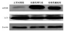
目的 探讨雷帕霉素靶蛋白/自噬(mTOR/自噬)信号通路在结核性气管支气管狭窄中的表达及作用。方法: 收集湖南省胸科医院2020年6月至2021年6月经外科评估需要手术切除病灶的肺曲菌患者,共10例,作为正常对照组。收集同期结核性气管支气管狭窄患者27例,包括13例结核性增生患者(结核性增生组),14例结核性瘢痕患者(结核性瘢痕组)。收集各组患者气管支气管组织,运用免疫组化检测mTOR、自噬相关蛋白LC3、转化生长因子-β1(TGF-β1)、胶原蛋白1(COL-1)蛋白表达情况,采用蛋白质印迹法检测mTOR、LC3蛋白表达情况,运用PCR检测TGF-β1、COL-1 mRNA的表达水平。结果: (1)结核性增生组、结核性瘢痕组中mTOR蛋白在免疫组化[分别为(0.074±0.008)和(0.041±0.004)]及蛋白质印迹法[分别为(3.397±0.312)和(2.261±0.175)]中的表达量均明显高于正常对照组[分别为(0.028±0.004)和(0.980±0.091)],差异均有统计学意义(t=0.045,P<0.001;t=0.013,P=0.036;t=2.419,P<0.001;t=1.283,P<0.001)。(2) 结核性增生组、结核性瘢痕组中TGF-β1蛋白在免疫组化[分别为(0.062±0.008)和(0.039±0.006)]及mRNA[分别为(6.930±0.606)和(3.350±0.582)]中的表达量均明显高于正常对照组[分别为(0.019±0.006)和(1.000±0.000)],差异均有统计学意义(t=0.043,P<0.001;t=0.020,P=0.009;t=5.930,P<0.001;t=2.353,P=0.001)。(3) 结核性增生组、结核性瘢痕组中COL-1蛋白在免疫组化[分别为(0.056±0.009)和(0.032±0.003)]及mRNA[分别为(6.803±1.110)和(2.730±0.547)]中的表达明显高于正常对照组[分别为(0.018±0.002)和(1.000±0.000)],差异均有统计学意义(t=0.038,P<0.001;t=0.013,P=0.026;t=5.803,P<0.001;t=1.730,P=0.025)。(4)结核性增生组、结核性瘢痕组中LC3蛋白在免疫组化[分别为(0.023±0.007)和(0.046±0.008)]及蛋白质印迹法[分别为(0.140±0.0303)和(0.236±0.030)]中的表达量均明显低于正常对照组[分别为(0.070±0.005)和(0.320±0.049)],差异均有统计学意义(t=0.047,P<0.001;t=0.023,P=0.009;t=0.179,P<0.001;t=0.083,P=0.034)。结论: 结核性气管支气管狭窄的发病可能与mTOR/自噬信号通路激活后TGF-β1和COL-1过度表达所致纤维化相关。

关键词: 结核, 支气管, 自噬, 纤维化

Abstract: Objective: To investigate the expression and role of rapamycin target protein/autophagy (mTOR/autophagy) signaling pathway in tuberculous tracheobronchial stenosis.Methods: A total of 10 patients with pulmonary Aspergillus who needed surgical resection in Hu’nan Chest Hospital from June 2020 to June 2021 were collected as the normal control group. And 27 patients with tuberculous tracheal bronchial stenosis over the same period were collected, including 13 patients with tuberculous hyperplasia (tuberculous hyperplasia group) and 14 patients with tuberculous scar (tuberculous scar group). The tracheal bronchial tissue of each group was collected. Immunohistochemistry was used to detect mTOR, autophagy-related protein LC3, transform growth factor-β1 (TGF-β1), collagen 1 (COL-1) protein expression, Western blotting was used to detect mTOR and the expression of LC3 prote, and PCR was used to detect the expression of TGF-β1 and COL-1 mRNA.Results: (1) By immunohistochemistry and western blotting, mTOR protein expressions in tuberculous hyperplasia group and tuberculous scar group were significantly higher than those in the normal control group ((0.074±0.008) vs. (0.028±0.004), t=0.045, P<0.001; (3.397±0.312) vs. (0.028±0.004), t=2.419, P<0.001; (0.041±0.004) vs. (0.980±0.091), t=0.013, P=0.036; (2.261±0.175) vs. (0.980±0.091), t=1.283, P<0.001). (2) By immunohistochemistry and and mRNA, TGF-β1 protein expressions in tuberculous hyperplasia group,and the tuberculous scar group were (0.062±0.008) and (0.039±0.006), (6.930±0.606) and (3.350±0.582), respectively; which were significantly higher than those in the normal control group (immunohistochemistry, 0.019±0.006, t=0.043, P<0.001 and t=0.020, P=0.009, respectively; mRNA, 1.000±0.000, t=5.930, P<0.001 and t=2.353, P=0.001, respectively). (3) In tuberculous hyperplasia group and the tuberculous scar group, COL-1 protein expressions were (0.056±0.009) and (0.032±0.003) by immunohistochemistry and (6.803±1.110) and (2.730±0.547) by mRNA, which were significantly higher than those in the normal control group ((0.018±0.002) and (1.000±0.000), t=0.038, P<0.001; t=0.013, P=0.026; t=0.013, P=0.026; t=5.803, P<0.001; t=1.730, P=0.025, respectively). (4) By immunohistochemistry and Western blotting, LC3 protein expressions in the tuberculous hyperplasia group andin the tuberculous scar group were (0.023±0.007) and (0.046±0.008), (0.140±0.030) and (0.236±0.030), which were significantly lower than those in the normal control group ((0.070±0.005) and (0.320±0.049), t=0.047, P<0.001; t=0.023, P=0.009; t=0.179, P<0.001; t=0.083, P=0.034).Conclusion: Tuberculous tracheobronchial stenosis may be related to fibrosis caused by overexpression of TGF-β1 and COL-1 after activation of mTOR/autophagy signaling pathway.

Key words: Tuberculous, Bronchi, Autophagy, Fibrosis

中图分类号: