结核与肺部疾病杂志 ›› 2025, Vol. 6 ›› Issue (1): 55-60.doi: 10.19983/j.issn.2096-8493.2024145

• 论著 • 上一篇    下一篇

超声引导下微波消融技术在局限性结核病变治疗中的价值分析

阎庆虎1, 薛峰2, 于泳3, 秦毅1, 阎庆梅4, 崔嘉1()   

  1. 1山东大学附属公共卫生临床中心/山东省公共卫生临床中心超声科,济南 250013
    2山东大学齐鲁医院放射科,济南 250012
    3山东第一医科大学附属省立医院超声科,济南 250000
    4济南市市中区人民医院急诊科,济南 250000
  • 收稿日期:2024-09-29 出版日期:2025-02-20 发布日期:2025-02-20
  • 通信作者: 崔嘉 E-mail:584872500@qq.com
  • 基金资助:
    山东省医药卫生科技项目(面上项目)(202309020993)

The value of ultrasound-guided microwave ablation in the treatment of localized tuberculous lesions

Yan Qinghu1, Xue Feng2, Yu Yong3, Qin Yi1, Yan Qingmei4, Cui Jia1()   

  1. 1Ultrasound Department of Shandong Public Health Clinical Center, Ji’nan 250013, China
    2Department of Radiology, Qilu Hospital, Shandong University, Ji’nan 250012, China
    3Ultrasound Department of Shandong First Medical University Affiliated Provincial Hospital, Ji’nan 250000, China
    4Emergency Department of Shizhong District People’s Hospital, Ji’nan 250000, China
  • Received:2024-09-29 Online:2025-02-20 Published:2025-02-20
  • Contact: Cui Jia E-mail:584872500@qq.com
  • Supported by:
    Shandong Province Medical and Health Science and Technology Project (General Project)(202309020993)

摘要:

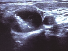
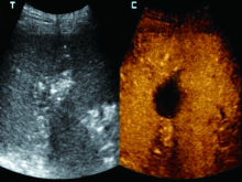
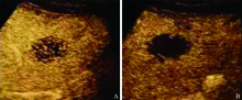
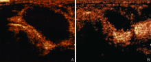
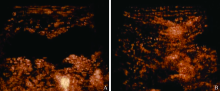
目的:探讨在常规内科结核药物治疗的基础上,利用超声引导下微波消融技术在局限性结核病变治疗中的应用价值。方法:回顾性分析2020年3月至2023年1月在山东省公共卫生临床中心和山东第一医科大学附属省立医院行超声引导下微波消融治疗的12例局限性结核病变患者,包括淋巴结结核6例、胸壁结核4例、肝脏结核2例。所有患者的局限性结核灶在微波消融后,行超声造影检查,消融后,3、6、9、12、24个月进行随访,测量消融灶体积,统计消融灶体积缩小率,评价消融效果。结果:消融术后,12例局限性结核患者的15个结核病灶超声造影均显示完全消融,术后3、6、9、12、24个月进行随访,消融灶体积分别为2.82(1.62,5.85)cm3、1.95(1.54,4.59)cm3、1.62(0.15,4.02)cm3、1.52(0.00,3.98)cm3、0.00(0.00,1.64)cm3,消融灶缩小率分别为51(36,59)%、62(52,72)%、69(58,97)%、71(62,100)%、100(72,100)%, 8个消融灶随访期内消失。术后24个月消融灶缩小率与术后3、6、9、12个月比较,差异均有统计学意义(χ2值分别为-3.41、-3.18、-3.06、-2.52,P值分别为0.001、0.001、0.002、0.012)。术中及术后所有患者均未出现周围脏器损伤、针道种植及结核播散等并发症,随访期未发现活动性进展或复发。结论:超声引导下微波消融治疗局限性结核病变具有较高的价值,值得临床推广。

关键词: 结核, 超声检查, 介入性, 治疗应用

Abstract:

Objective: This study aims to evaluate the clinical efficacy and potential applications of ultrasound-guided microwave ablation as an adjunct to conventional anti-tuberculosis pharmacotherapy in the management of localized tuberculous lesions. Methods: A retrospective analysis was conducted on 12 patients with localized tuberculous lesions who underwent ultrasound-guided microwave ablation at the Shandong Public Health Clinical Center and the Shandong First Medical University Affiliated Provincial Hospital between March 2020 and January 2023. The cohort included 6 cases of lymph node tuberculosis, 4 cases of chest wall tuberculosis, and 2 cases of liver tuberculosis. Contrast-enhanced ultrasound (CEUS) was performed post-ablation for all patients to evaluate treatment efficacy. Follow-up assessments were carried out at 3, 6, 9, 12, and 24 months post-ablation to measure the ablation lesion volume and calculate its reduction rate. Data were expressed as median (interquartile range, M(P25, P75)) and analyzed using the Z-test to assess the effectiveness of the ablation procedure. Results: Contrast-enhanced ultrasound confirmed complete ablation in all 15 localized tuberculous lesions across 12 patients immediately after the procedure. During follow-up at 3, 6, 9, 12, and 24 months post-ablation, the median lesion volumes were recorded as 2.82 (1.62,5.85) cm3, 1.95 (1.54,4.59) cm3, 1.62 (0.15,4.02)cm3, 1.52 (0.00,3.98)cm3, 0.00 (0.00,1.64)cm3, respectively. Correspondingly, the shrinkage rates of the ablation lesions were 51 (36,59)%, 62 (52,72)%, 69 (58,97)%, 71 (62,100)%, 100 (72,100)%. By the end of the follow-up period, 8 ablation lesions had completely resolved. The reduction rate of ablation lesions at 24 months post-surgery was significantly higher compared to those at 3, 6, 9, and 12 months, with corresponding χ2 values of -3.41, -3.18, -3.06, and -2.52 (P=0.001, 0.001, 0.002, and 0.012, respectively). No complications, such as peripheral organ damage, needle track implantation, or tuberculosis dissemination, were observed during or after the procedure. Furthermore, no active disease progression or recurrence was detected throughout the follow-up period. Conclusion: Ultrasound-guided microwave ablation demonstrates significant efficacy and safety in the treatment of localized tuberculous lesions, highlighting its potential as a valuable minimally invasive therapeutic option deserving broader clinical application.

Key words: Tuberculosis, Ultrasonography, interventional, Therapeutic uses

中图分类号: