结核病与肺部健康杂志 ›› 2018, Vol. 7 ›› Issue (4): 245-250.doi: 10.3969/j.issn.2095-3755.2018.04.005

• 论著 • 上一篇    下一篇

磨玻璃样肺腺癌多维度CT征象特点对其浸润程度预测的临床研究

李凤,邱太春,黎佳维,伍建林()   

  1. 116001 大连大学附属中山医院放射科
  • 收稿日期:2018-10-22 出版日期:2018-12-30 发布日期:2019-01-08
  • 通信作者: 伍建林 E-mail:wujianlin@vip.163.com

Clinical study on multi-dimensional CT signs of ground glass lung adenocarcinoma to predict its invasion degree

Feng LI,Tai-chun QIU,Jia-wei LI,Jian-lin WU()   

  1. Department of Radiology, Zhongshan Hospital Affiliated to Dalian University, Dalian 116001, China
  • Received:2018-10-22 Online:2018-12-30 Published:2019-01-08
  • Contact: Jian-lin WU E-mail:wujianlin@vip.163.com

摘要:

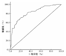
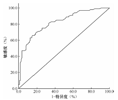
目的 探讨多维度CT征象对以磨玻璃样结节(GGN)为特征的肺腺癌浸润程度或病理亚型进行预测的临床应用价值。方法 回顾性分析2013年1月至2017年12月大连大学附属中山医院经病理证实230例肺磨玻璃样结节患者的CT扫描资料,其中12例患者胸部CT扫描显示肺部有2个磨玻璃样结节。按照2011版肺腺癌最新分类标准,分为浸润前病变110个(浸润前病变组)、微浸润腺癌32个(微浸润腺癌组)及浸润性腺癌100个(浸润性腺癌组)。分析CT征象包括其一般维度、边缘维度、内部维度及管腔维度(血管征象分型及支气管征象分型)。结果 (1)一般维度征象,病灶形态为圆形或类圆形在浸润前病变组、微浸润腺癌组和浸润性腺癌组的发生率分别为59.1%(65/110)、68.8%(22/32)、55.0%(55/100),差异无统计学意义(χ 2=1.904,P>0.05);3组GGN病灶直径中,浸润性病变组[(17.4±7.0)mm]>微浸润病变组[(11.0±5.1)mm]>浸润前病变组[(10.0±4.7)mm],差异有统计学意义(U=68.312,P<0.05);ROC曲线分析显示,鉴别浸润前病变与浸润性腺癌病变(包括微浸润与浸润性病变组)的临界值为1.29cm,诊断敏感度为70.0%,特异度为80.7%,ROC曲线下面积(AUC)为0.802。(2)边缘维度征象,分叶征在浸润前病变组、微浸润腺癌组和浸润性腺癌组的发生率分别为21.8%(24/110)、56.3%(18/32)、82.0%(82/100),差异有统计学意义(χ 2=76.304,P<0.05)。(3)内部维度征象,浸润前病变组、微浸润腺癌组和浸润性腺癌组空泡征的发生率分别为10.9%(12/110)、18.8%(6/32)、55.0%(55/100),差异有统计学意义(χ 2=50.620,P<0.05)。(4)管腔维度征象,血管征象分型在浸润性腺癌以Ⅲ型(57.0%,57/100)、Ⅳ型(40.0%,40/100)多见,微浸润腺癌Ⅲ型(62.5%,20/32)多见,浸润前病变Ⅱ型(65.5%,72/110)多见;而支气管征象分型在浸润性腺癌以Ⅱ型(34.0%,34/100)、Ⅲ型(37.0%,37/100)多见,微浸润腺癌多见Ⅲ型(40.6%,13/32),浸润前病变多见Ⅳ型(50.9%,56/110),3组间差异均有统计学意义(χ 2值分别为141.749、134.268,P值均<0.05);将血管征象与支气管征象分型单独或联合进行浸润前病变与浸润性腺癌预测的准确率分别为86.4%(209/242)、82.2%(199/242)和89.3%(216/242)。 结论 通过多维度CT征象综合分析有助于对GGN为特征的肺腺癌浸润程度及病理亚型术前预测,尤其将血管征象及支气管征象分型同时出现且联合诊断时的鉴别效能高于各征象单独诊断,为临床术前预测GGN为特征的肺腺癌浸润程度提供了新的信息与方向。

关键词: 腺癌, 体层摄影术,X线计算机, 诊断显像, 磨玻璃样结节

Abstract:

Objective Identify the value of multi-dimensional CT sings in predicting the tumor invasion degree and pathological subtype of lung adenocarcinoma characterized with ground-glass nodule(GGN).Methods Retrospective analysis of CT imaging data of 230 patients with pulmonary ground-glass nodules confirmed by pathology from January 2013 to December 2017 in Zhongshan Hospital affiliated to Dalian University, 12 patients had 2 lung ground-glass nodules. According to the latest classification criteria for lung adenocarcinoma in 2011, there were 110 preinvasive lesions, 32 microinvasive and 100 invasive adenocarcinomas. Analysis of CT signs, including general dimensions, marginal dimensions, internal dimensions, and lumen dimensions (including vascular and bronchial CT signs)Results (1) In general dimension signs, the proportion of round/quasi-round lesions in the preinvasive lesion group, microinvasive adenocarcinoma group and invasive adenocarcinoma group were 59.1% (65/110), 68.8% (22/32), 55.0% (55/100) respectively, the difference was not statistically significant (χ 2=1.904,P>0.05).However, the diameters of GGN lesions in three groups were (10.0±4.7)mm, (11.0±5.1)mm and (17.4±7.0)mm respectively,the difference was significant (U=68.312,P<0.05). ROC curve analysis showed that the preinvasive lesions and invasive adenocarcinoma lesions (including microinvasive and invasive lesions) had a critical value of 1.29 cm, and the diagnostic sensitivity is 70.0%, the specificity is 80.7%, and the AUC (area under ROC curve) is 0.802.(2)In marginal dimension signs, the proportion of lobulated signs were 21.8% (24/110), 56.3% (18/32) and 82.0% (82/100) in preinvasive lesion group, microinvasive adenocarcinoma group and invasive adenocarcinoma group respectively. The difference was statistically significant (χ 2=76.304,P<0.05).(3) In internal dimension signs, the proportion of vacuole signs in three groups were 10.9% (12/110), 18.8% (6/32), 55.0% (55/100) respectively. The difference was statistically significant(χ 2=50.620,P<0.05). (4) In lumen dimension signs, Vascular signs were obvious in type Ⅲ (57.0%, 57/100) and Ⅳ (40.0%, 40/100) invasive adenocarcinoma, type Ⅲ (62.5%, 20/32) microinvasive adenocarcinoma and in type Ⅱ (65.5%, 72/110) preinvasive lesions group. Bronchial signs were obvious in invasive adenocarcinoma type Ⅱ (34.0%, 34/100) and Ⅲ (37.0%, 37/100), microinvasive adenocarcinoma type Ⅲ (40.6%, 13/32) and preinvasive lesions type Ⅳ (50.9%, 56/110). There were significant differences among the three groups(χ 2 values are 141.749、134.268,P<0.05).The accuracy of predicting probability of preinvasive lesions and invasive adenocarcinoma through vascular signs and bronchial signs alone or in combination were 86.4%(209/242)、82.2%(199/242)、89.3%(216/242)respectively. Conclusion Comprehensive analysis of multi-dimensional CT signs is helpful for preoperative prediction of GGN-like lung adenocarcinoma infiltration and pathological subtypes, especially the simultaneous identification of vascular signs and bronchial signs and combined diagnosis were more effective than the diagnosis of each sign. Multi-dimensional CT signs provides new information and direction for the preoperative prediction of GGN-like lung adenocarcinoma infiltration.

Key words: Adenocarcinoma, Tomography, X-ray computed, Diagnostic imaging, Ground-glass nodule (GGN)