结核与肺部疾病杂志 ›› 2023, Vol. 4 ›› Issue (4): 301-307.doi: 10.19983/j.issn.2096-8493.20230061

• 论著 • 上一篇    下一篇

支气管镜下肺癌病理类型及影像学特征分析

王瑶, 刘敏, 胡智敏, 余盼丽, 吴鸣镝, 靖秋生()   

  1. 武汉市肺科医院/武汉市结核病防治所内镜中心,武汉 430030
  • 收稿日期:2023-04-21 出版日期:2023-08-20 发布日期:2023-08-15
  • 通信作者: 靖秋生,Email: 1804531120@qq.com
  • 作者简介:注:刘敏与王瑶对本研究具有同等贡献,为并列第一作者

Analysis of pathological types and imaging features of lung cancer under bronchoscopy

Wang Yao, Liu Min, Hu Zhimin, Yu Panli, Wu Mingdi, Jing Qiusheng()   

  1. Endoscopy Center, Wuhan Pulmonary Hospital/Wuhan Institute for Tuberculosis Prevention and Control, Wuhan 430030, China
  • Received:2023-04-21 Online:2023-08-20 Published:2023-08-15
  • Contact: Jing Qiusheng, Email: 1804531120@qq.com

摘要:

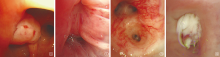
目的: 分析肺癌患者电子支气管镜下影像学特征及病理类型,探讨其辅助诊断价值。 方法: 采用回顾性分析方法,收集2020年7月至2022年6月武汉市肺科医院内镜中心行电子支气管镜检查并获得病理标本且明确诊断为肺癌的213例患者临床资料。分析患者性别、年龄、镜下病灶特征和部位、活检钳取样块数在不同肺癌病理类型中的差异。 结果: 213例肺癌患者中,肺鳞癌108例(50.7%)、肺腺癌51例(23.9%)、肺小细胞癌24例(11.3%)、其他类型或不能明确病理分型的恶性肿瘤30例(14.1%),其中2例男性患者为肺鳞癌并发肺小细胞癌。肺癌患者以男性多见[85.4%(182/213)],且鳞癌多见于男性[98.1%(106/108)],而女性中腺癌的占比较高[64.5%(20/31)],不同性别的不同病理类型占比的差异有统计学意义(χ2=48.293,P=0.000)。病变累及部位:右侧支气管[59.2%(129例)]>左侧支气管[42.7%(91例)]>主气管[16.9%(36例)]。鳞癌以增生型表现为主(66.7%,72/108),在镜下多表现为新生物向管腔内生长,常合并糜烂、溃疡或附着白色坏死物(36.1%,39/108);而58.8%(30/51)的腺癌和70.8%(17/24)的小细胞癌在镜下多为浸润表现,表现为沿管壁浸润性生长,病灶处黏膜充血、肿胀、肥厚、粗糙或间嵴增宽,并分别有41.2%(21/51)的腺癌和87.5%(21/24)的小细胞癌病灶处伴有血管密集、增粗、紊乱、迂曲等血管异常,不同病理类型在镜下影像学表现的差异有统计学意义(χ2=31.113,P=0.000)。用于病理诊断需钳取活检组织数以3块及以下为主(62.0%,132/213),仅6.6%(14/213)的肺癌患者需要6块以上。 结论: 支气管镜检查可直接观察病灶部位及影像特征表现,有助于镜下判断肺癌的病理类型,并可直接取活检组织进行病理学诊断,是发现并诊断肺癌的重要方式。

关键词: 癌,非小细胞肺, 支气管镜检查, 疾病特征

Abstract:

Objective: To analyze the pathological types and imaging characteristics of lung cancer under electronic bronchoscopy and its diagnostic value. Methods: A retrospective analysis was conducted to collect the clinical data of 213 patients who underwent electronic bronchoscopy in the Endoscopy Center of Wuhan Pulmonary Hospital from July 2020 to June 2022. Gender, age, microscopic lesion characteristics and location, number of biopsy forceps samples and pathological type of lung cancer were analyzed. Results: All of 213 cases of lung cancer, 108 cases (50.7%) had squamous cell carcinoma, 51 cases (23.9%) had lung adenocarcinoma, 24 cases (11.3%) had lung small cell carcinoma, and 30 cases (14.1%) had malignant tumor of other types or could not be clearly classified, among them 2 male patients had lung squamous cell carcinoma complicated with lung small cell carcinoma. Among the 213 cases of lung cancer, the majority of them were male (85.4%, 182/213). Squamous cell carcinoma was mostly found in male (98.1%, 106/108) and adenocarcinoma in female (64.5%, 20/31), there was a significant difference of pathological types between the two genders (χ2=48.293, P=0.000). The most frequent sites of the lesions were: right branch (59.2%, 126 cases) >left branch (42.7%, 91 cases) >main trachea (16.9%, 36 cases). The squamous cell carcinoma was mainly hyperplastic (66.7%, 72/108), showing as new organism grew into the lumen under microscope, often accompanied by erosion, ulcer or white necrosis (36.1%, 39/108); 58.8% (30/51) of adenocarcinoma and 70.8% (17/24) of small cell carcinoma were found to be infiltrated under microscope. There were 41.2% (21/51) of adenocarcinoma and 87.5% (21/24) of small cell carcinoma getting lesions with vascular abnormalities, such as dense, thickened, disordered and tortuous vessels. There was a significant difference in the microscopic manifestations of different pathological types of lung cancer (χ2=31.113, P=0.000). 62.0% (132/213) patients with lung cancer required 3 pieces or less biopsy tissues for pathological diagnosis,and only 6.6% (14/213) of lung cancer patients needed more than 6 pieces. Conclusion: Bronchoscopy could directly observe the lesion and its imaging manifestations which could be of help to determine the pathological type of lung cancer, also could directly take the biopsy tissue for pathological diagnosis. Therefore, it is an important way to detect and diagnose lung cancer.

Key words: Carcinoma, non-small-cell lung, Bronchoscopy, Disease attributes

中图分类号: