结核与肺部疾病杂志 ›› 2022, Vol. 3 ›› Issue (5): 388-393.doi: 10.19983/j.issn.2096-8493.20220117

• 论著 • 上一篇    下一篇

人造石相关矽肺合并肺结核与非结核分枝杆菌肺病28例临床分析

陈子丹, 毛翎()   

  1. 同济大学附属上海市肺科医院,上海 200433
  • 收稿日期:2022-07-20 出版日期:2022-10-20 发布日期:2022-10-14
  • 通信作者: 毛翎 E-mail:maoling113@sina.com
  • 基金资助:
    2022上海市卫生健康地方标准预研制项目(2022WB06)

Clinical analysis of 28 cases of artificial stone associated silicosis complicated with pulmonary tuberculosis and non-tuberculous mycobacteria disease

Chen Zidan, Mao Ling()   

  1. Shanghai Pulmonary Hospital, Tongji University School of Medicine, Shanghai 200433, China
  • Received:2022-07-20 Online:2022-10-20 Published:2022-10-14
  • Contact: Mao Ling E-mail:maoling113@sina.com
  • Supported by:
    Shanghai Local Health Standard Pre-Development Project(2022WB06)

摘要:

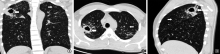
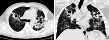
目的: 了解人造石(artificial stone,AS)相关矽肺(简称“AS-矽肺”)合并肺结核与非结核分枝杆菌(NTM)肺病患者的临床特征,为临床诊断提供参考依据。方法: 采用回顾性分析的方法,以2017年1月至2020年12月上海市肺科医院尘肺科收治的确诊AS-矽肺合并肺结核与NTM肺病的28例患者为研究对象,观察和记录患者临床症状、病原学检查结果、胸部CT影像学特征及治疗转归。结果: 28例患者中,AS-矽肺合并NTM肺病患者和合并肺结核患者均为14例,各占50.0%,主要临床表现为咳嗽(92.9%,26/28)、咳痰(89.3%,25/28)、气喘(64.3%,18/28)。AS-矽肺合并NTM肺病患者涂片阳性率为64.3%(9/14),高于合并肺结核的患者(28.6%,4/14);免疫学检查阳性率为25.0%(3/12),低于合并肺结核的患者(40.0%,4/10)。28例患者胸部CT主要表现为磨玻璃影(53.6%,15/28)、小结节影(92.9%,26/28)、斑片影(25.0%,7/28)、团块影(71.4%,20/28)、空洞(85.7%,24/28);AS-矽肺合并NTM肺病患者空洞以洞壁光滑(70.0%,14/20)和薄壁空洞(55.0%,11/20)为主,合并肺结核患者上述征象占比均为0.0%(0/14)。AS-矽肺合并NTM肺病患者和合并肺结核患者治疗有效率分别为2/8和50.0%(6/12)。结论: AS-矽肺合并肺结核与合并NTM肺病患者治疗有效率较低。影像学差异可为临床诊断提供依据,当出现洞壁光滑的薄壁空洞时警惕可能合并NTM肺病。

关键词: 矽肺, 结核, 分枝杆菌,非典型性, 治疗结果

Abstract:

Objective: To investigate the clinical characteristics of artificial stone associated silicosis (AS-silicosis) complicated with pulmonary tuberculosis and non-tuberculous mycobacteria disease, and to provide reference for clinical diagnosis. Methods: A retrospective study was conducted, 28 patients with AS-silicosis complicated with pulmonary tuberculosis and non-tuberculous mycobacteria disease admitted to the Department of Pneumoconiosis of Shanghai Pulmonary Hospital from January 2017 to December 2020 were selected as study objects. The clinical symptoms, bacteriological test results, Mycobacterium tuberculosis (MTB) nucleic acid test results, chest CT imaging features and treatment outcomes of all subjects were collected and analyzed. Results: Among all 28 patients, AS-silicosis combined with non-tuberculous mycobacterium (NTM) lung disease and with pulmonary tuberculosis were both 14, accounting for 50.0% for each. The main clinical manifestations of all subjects were cough (92.9%, 26/28), expectoration (89.3%, 25/28), and puff (64.3%, 18/28). Smear positive rate among patients with NTM (64.3%, 9/14) was higher than those with pulmonary tuberculosis (28.6%, 4/14), while immunological test positive rate among NTM patients (25.0%, 3/12) was lower than those with pulmonary tuberculosis (40.0%, 4/10). Ground glass opacity (53.6%, 15/28), small nodule (92.9%, 26/28), patchy (25.0%, 7/28), progressive mass fibrosis (PMF)(71.4%, 20/28) and cavity (85.7%, 24/28) were the main manifestations of CT images in all subjects, and there was no significant difference between patients with NTM and patients with pulmonary tuberculosis. Cavities of patients with NTM mainly showed having thin wall (55.0%, 11/20) with smooth inner wall (70.0%, 14/20), while 0.0% (0/14) of cavities in patients with pulmonary tuberculosis got this feature. If imaging improvement such as CT lesion shrinking, cavity narrowing or close were considered as the necessary conditions for effective treatment, the effective treatment rate in patients with NTM and with pulmonary tuberculosis were 2/8 and 50.0% (6/12), respectively. Conclusion: AS-silicosis combined with pulmonary tuberculosis and non-tuberculous mycobacteria disease have low effective treatment rate. The difference of CT images could provide a basis for clinical diagnosis as thin-walled cavity with smooth inner wall may be associated with NTM lung disease.

Key words: Silicosis, Tuberculosis, Mycobacteria, atypical, Treatment outcome

中图分类号: