结核病与肺部健康杂志 ›› 2018, Vol. 7 ›› Issue (4): 241-244.doi: 10.3969/j.issn.2095-3755.2018.04.004

• 论著 • 上一篇    下一篇

囊腔型肺癌与空洞型肺结核的CT鉴别诊断研究

谭洋,邱太春,伍建林(),张冠男,张国庆   

  1. 116001 大连大学附属中山医院放射科(谭洋、邱太春、伍建林);大连市结核病医院放射科(张冠男、张国庆)
  • 收稿日期:2018-10-22 出版日期:2018-12-30 发布日期:2019-01-08
  • 通信作者: 伍建林 E-mail:wujianlin@vip.163.com

Differential diagnosis of lung cancers associated with cystic airspaces and cavitary pulmonary tuberculosis by CT

Yang TAN,Tai-chun QIU,Jian-lin WU(),Guan-nan ZHANG,Guo-qing ZHANG   

  1. Department of Radiology, Affiliated Zhongshan Hospital of Dalian University, Dalian 116001, China
  • Received:2018-10-22 Online:2018-12-30 Published:2019-01-08
  • Contact: Jian-lin WU E-mail:wujianlin@vip.163.com

摘要:

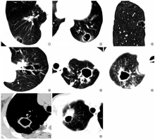
目的 回顾性分析囊腔型肺癌与空洞型肺结核的CT表现特点,提高两者的诊断与鉴别诊断水平。方法 选取2015年3月至2018年5月于大连大学附属中山医院收治并经手术和(或)穿刺活检病理证实的23例囊腔型肺癌患者作为肺癌组;选取同期大连市结核病医院收治的经痰细菌学检查或临床治疗随访证实的31例空洞型肺结核患者作为结核组。收集患者的CT检查资料,对两组研究对象CT征象进行比较和分析。结果 肺癌组(60.9%,14/23)和结核组(61.3%,19/31)患者病灶均以右肺好发;肺癌组病灶直径平均为(17.19±11.02)mm,明显小于结核组的(27.13±10.42)mm,差异有统计学意义(t=-2.55,P=0.014)。肺癌组囊腔平均壁厚为(1.91±0.73)mm,明显小于结核组的(2.69±0.47)mm,差异有统计学意义(t=3.05,P<0.05)。肺癌组囊腔内见壁结节者占47.8%(11/23),高于结核组空洞内见壁结节者(19.4%,6/31),差异有统计学意义(χ 2=7.69,P<0.05)。肺癌组囊腔内见血管分支影者占43.5%(10/23),而结核组患者空洞内均未见血管分支影,差异有统计学意义(χ 2=11.71,P<0.01)。肺癌组囊腔边缘可见分叶征者5例(21.7%),见毛刺征者13例(56.5%);结核组空洞边缘可见分叶征者6例(19.4%),见毛刺征者16例(51.6%),两组间差异均无统计学意义(χ 2=0.61,P>0.05)。肺癌组出现胸膜凹陷征者12例(52.2%),结核组出现23例(74.2%)并发不同程度邻近胸膜增厚,两组间差异有统计学意义(χ 2=11.04,P<0.05)。结论 囊腔型肺癌的CT表现具有一定特征性,典型者可在临床上藉此做出该类肺癌的提示性诊断,并有助于与空洞型肺结核进行鉴别。

关键词: 肺肿瘤, 结核,肺, 体层摄影术,X线计算机, 诊断,鉴别

Abstract:

Objective To retrospectively analyze the CT features of lung cancers associated with cystic airspaces and cavitary pulmonary tuberculosis, and to improve the diagnosis and differential diagnosis.Methods Twenty-three cases with lung cancer associated with cystic airspaces who were admitted to Affiliated Zhongshan Hospital of Dalian University and confirmed by surgery and/or biopsy between March 2015 and May 2018 were collected as lung cancer group. In the same period, 31 cases of cavitary tuberculosis confirmed by laboratory bacteriological examination and clinical follow-up or treatment from Tuberculosis Hospital of Dalian were selected as tuberculosis group. The CT data of the patients were collected and the CT feature were compared between two groups.Results Most of the lesions were located in the right lung (lung cancer group: 60.9% (14/23); tuberculosis group: 61.3% (19/31)). The lesion size of lung cancer group was smaller than that of the tuberculosis group ((17.19±11.02) mm vs (27.13±10.42) mm), and the difference was statistically significant (t=-2.55, P=0.014). The average thickness of cavity wall of the lung cancer group ((1.91±0.73) mm) was smaller than that of the tuberculosis group ((2.69±0.47) mm), and the difference was statistically significant (t=3.05, P<0.05). The rate of patients with wall nodules in the cystic cavity in the lung cancer group (47.8%, 11/23) was higher than that in the tuberculosis group (19.4%, 6/31), and the difference was statistically significant (χ 2=7.69, P<0.05). In the lung cancer group, shadow of the vascular branch in the cystic cavity was shown in 43.5% (10/23) of the patients, but no vascular branch shadow appeared in the cavity of the tuberculosis group. The difference was statistically significant (χ 2=11.71, P<0.01). In the lung cancer group, 5 patients (21.7%) had lobular stenosis at the edge of the cyst, and 13 patients (56.5%) had burr marks. In the tuberculosis group, the lobes were visible at the margin of the cavity in 6 patients (19.4%), and 16 patients (51.6%) had burr sign. The difference was not statistically significant (χ 2=0.61, P>0.05). Among the patients in the lung cancer group, 12 patients (52.2%) had pleural indentation, and 23 patients (74.2%) in the tuberculosis group had different degrees of adjacent pleural thickening. The difference was statistically significant (χ 2=11.04, P<0.05). Conclusion CT findings of lung cancers associated with cystic airspaces are characteristic. Typical CT features can help us make a suggestive diagnosis of this type of lung cancer, and help to differential diagnosis from cavitary tuberculosis.

Key words: Lung neoplasms, Tuberculosis, pulmonary, Tomography, X-ray computed, Diagnosis, differential