结核病与肺部健康杂志 ›› 2018, Vol. 7 ›› Issue (4): 255-260.doi: 10.3969/j.issn.2095-3755.2018.04.007

• 论著 • 上一篇    下一篇

以肾自截为主要表现的肾结核二例并文献复习

张洁,刘玉琴(),李雨泽,韩立清,刘淑芹,李宏明,孙洋,齐玉玲   

  1. 150500 哈尔滨,黑龙江省传染病防治院(张洁、刘玉琴、李雨泽、韩立清、李宏明、孙洋、齐玉玲);牡丹江医学院第二附属医院(刘淑芹)
  • 收稿日期:2018-11-21 出版日期:2018-12-30 发布日期:2019-01-08
  • 通信作者: 刘玉琴 E-mail:liuyuqin_ssy@163.com

Renal tuberculosis as manifestation of autonephrectomy in two cases and literature review

Jie ZHANG,Yu-qin LIU(),Yu-ze LI,Li-qing HAN,Shu-qin LIU,Hong-ming LI,Yang SUN,Yu-ling QI   

  1. Infectious Disease Hospital of Heilongjiang Province, Harbin 150500, China
  • Received:2018-11-21 Online:2018-12-30 Published:2019-01-08
  • Contact: Yu-qin LIU E-mail:liuyuqin_ssy@163.com

摘要:

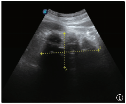
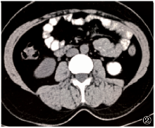
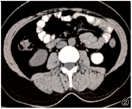
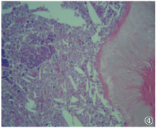
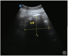
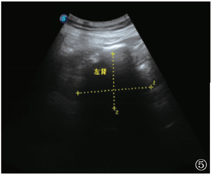
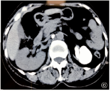
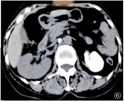
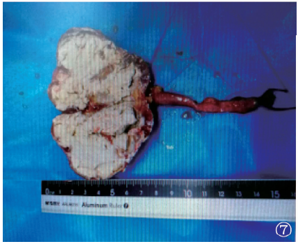
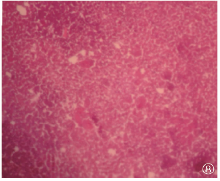
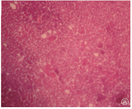
目的 总结肾自截的临床、影像及病理特点,以提高肾自截的临床诊断水平。方法 对黑龙江省传染病防治院2017年9月11日和2017年12月4日各收治的1例(共2例)肾自截患者的临床特点、病理表现、实验室检查结果、影像学检查结果,以及治疗及转归等情况进行分析。 并以2000—2018年为检索时间,以“肾结核”、“肾自截”为检索词,从PubMed、万方数据库、中国知网检索到10篇文献,入组患者12例;收集患者的一般资料、病程时间、病变部位、临床表现、实验室及影像学检查结果、并发症、诊断方法、治疗及转归情况,进行汇总分析。结果 2例患者均曾有尿频、尿急、尿痛、腰部疼痛症状,结核分枝杆菌分子生物学(尿液结核分枝杆菌耐药基因芯片法、血结核感染T细胞斑点试验)检测结果均为阳性;肾脏CT均表现为左肾体积缩小,完全钙化,静脉肾盂造影均为左肾盂肾盏及输尿管未显影。临床诊断为肾自截。2例患者均接受左肾切除,术后大体病理表现为肾组织切面灰黄囊性,囊内为灰黄灰白坏死灶、肾组织内可见淡黄色豆渣样油腻物。术后口服抗结核药物治疗(帕司烟肼或异烟肼、利福平、乙胺丁醇、吡嗪酰胺),疗程1年,分别于出院6、9个月时,尿频、尿急、尿痛症状明显减轻或消失。通过文献复习与筛选,共获得12例肾自截患者的临床资料,加上笔者报告的2例患者,共计14例。其中,男9例,女5例;年龄34~77岁;双侧肾结核3例,单侧肾结核11例(左肾6例,右肾5例);12例病变肾脏缩小伴钙化,2例病变肾脏增大,呈类圆形囊状,周边呈厚壁环状钙化;2例并发高血压病,1例并发精囊结核及膀胱结核,2例并发附睾结核,1例并发腹膜后淋巴结结核,1例并发右侧自发性肾瘘,3例并发对侧输尿管结石伴急性肾功能衰竭。6例患者手术切除病变肾脏,5例口服抗结核药物治疗,3例治疗方案不详。14例患者经治疗,尿频、尿急、尿痛症状均消失。结论肾自截临床症状较为严重,并发症较多,影像学检查是诊断的重要手段,尽早切除病变肾脏为治疗的重要方法之一。

关键词: 结核,肾, 超声检查, 体层摄影术,X线计算机, 病理状态,体征和症状, 综述文献(主题), 总结性报告(主题)

Abstract:

Objective To summarize the clinical, imaging and pathological features of renal tuberculosis as manifestation of autonephrectomy in order to improve the clinical diagnosis level of autonephrectomy.Methods We analyzed the clinical features, pathological manifestations, laboratory results, imaging characteristics, treatment and prognosis in two cases of renal tuberculosis with autonephrectomy admitted in Infectious Disease Hospital of Heilongjiang Province on Nov. 11, 2017 and Dec. 4, 2017, respectively. Taking 2000-2018 as the retrieval time and “renal tuberculosis, autonephrectomy” as the key word, 10 articles including 12 cases were retrieved from PubMed, Wanfang database and China National Knowledge Infrastructure (CNKI). The general information, duration of disease, location of lesion, clinical manifestations, laboratory and imaging results, complications, diagnostic methods, treatment and prognosis were collected and made meta-analysis.Results There were frequent micturition, urgent micturition, odynuria and lumbago in two cases. The results of molecular biology of Mycobacterium tuberculosis including the gene chip assay for Mycobacterium tuberculosis resistance in urine and T-SPOT.TB test in blood were all positive. Renal CT showed volumetric reduction and complete calcification in the left renal. Intravenous pyelography showed no development of left renal pelvis, renal calyx and ureter. Clinical diagnosis was autonephrectomy. Two patients underwent left nephrectomy. The gross pathological features were grayish yellow cysts on the cut surface of renal tissue, gray-yellow gray-white necrotic foci in the capsule, and yellowish soybean dregs like greasy substance in the renal tissue. Chemotherapy containing pasiniazide or isoniazid, rifampicin, ethambutol, pyrazinamide was performed after operation for one year. After 6 and 9 months of discharge, the symptoms of frequent micturition, urgent micturition, odynuria disappeared. There were 14 cases data including 12 cases with autonephrectomy from literature screening and 2 cases from the report of this study. Of 14 cases, 9 cases were male and 5 cases were female, aged from 34 to 77 years old. There were bilateral renal tuberculosis in 3 cases and unilateral renal tuberculosis in 11 cases including 6 cases in left renal and 5 cases in right renal. The volume of diseased renal reduced and complicated with calcification in 12 cases, and the volume of diseased renal enlarged with circular cysts and thick-walled circular calcification in 2 cases. There were complicated with hypertension in 2 cases, seminal vesicle tuberculosis and bladder tuberculosis in 1 case, epididymis tuberculosis in 2 cases, retroperitoneal lymph node tuberculosis in 1 case, right spontaneous renal fistula in 1 case, and contralateral ureteral calculi complicated with acute renal failure in 3 cases. Nephrectomy of diseased renal underwent in 6 cases, and chemotherapy was performed in 5 cases, and treatment was done with unknown regimen in 3 cases. The symptoms of frequent micturition, urgent micturition, odynuria disappeared in all cases after treatment.Conclusion The clinical symptoms are more serious with more complications in renal tuberculosis with nephrectomy. Imaging examination is an important means of diagnosis. Nephrectomy of diseased renal is one of the important methods of treatment.

Key words: Tuberculosis, renal, Ultrasonography, Tomography, X-ray computed, Pathological conditions, signs and symptoms, Review literature as topic, Consensus development conferences as topic