
Journal of Tuberculosis and Lung Disease ›› 2022, Vol. 3 ›› Issue (2): 137-141.doi: 10.19983/j.issn.2096-8493.20210135
Previous Articles Next Articles
ZHOU Lei, LUO Li, LU Zhi-bin, DING Yan, XIAO Yang-bao
Received:2021-10-21
Online:2022-06-30
Published:2022-04-18
Supported by:CLC Number:
ZHOU Lei, LUO Li, LU Zhi-bin, DING Yan, XIAO Yang-bao. Mechanism of rapamycin target protein/autophagy signaling pathway in tuberculous tracheal bronchial stenosis[J]. Journal of Tuberculosis and Lung Disease , 2022, 3(2): 137-141. doi: 10.19983/j.issn.2096-8493.20210135
Add to citation manager EndNote|Ris|BibTeX
URL: http://www.jtbld.cn/EN/10.19983/j.issn.2096-8493.20210135
| 蛋白名称 | 上游引物(5′-3′) | 下游引物(5′-3′) |
|---|---|---|
| TGF-β1 | AGCAACAATTCCTG- GCGATACCTC | CAATTTCCCCTCCA- CGGCTCA |
| COL-1 | GCAAGAACCCCGCCC- GCACC | GCTCTCGCCGAACC- AGACATGCC |
| β-actin | ACCCTGAAGTACCCC- ATCGAG | AGCACAGCCTGGAT- AGCAAC |
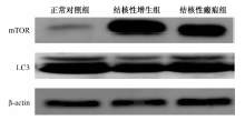
| 组别 | mTOR蛋白 | LC3蛋白 | TGF-β1蛋白 | COL-1蛋白 |
|---|---|---|---|---|
| 正常对照组(10例) | 0.028±0.004 | 0.070±0.005 | 0.019±0.006 | 0.018±0.002 |
| 结核性增生组(13例) | 0.074±0.008 | 0.023±0.007 | 0.062±0.008 | 0.056±0.009 |
| 结核性瘢痕组(14例) | 0.041±0.004 | 0.046±0.008 | 0.039±0.006 | 0.032±0.003 |
| F值 | 47.327 | 29.888 | 33.345 | 36.457 |
| P值 | <0.001 | 0.001 | 0.001 | <0.001 |
| t值a | 0.045 | 0.047 | 0.043 | 0.038 |
| P值 | <0.001 | <0.001 | <0.001 | <0.001 |
| t值b | 0.013 | 0.023 | 0.020 | 0.013 |
| P值 | 0.036 | 0.009 | 0.009 | 0.026 |
| t值c | 0.033 | 0.024 | 0.023 | 0.025 |
| P值 | 0.001 | 0.008 | 0.005 | 0.002 |

| 组别 | mTOR蛋白 | LC3蛋白 |
|---|---|---|
| 正常对照组(10例) | 0.980±0.091 | 0.320±0.049 |
| 结核性增生组(13例) | 3.397±0.312 | 0.140±0.030 |
| 结核性瘢痕组(14例) | 2.261±0.175 | 0.236±0.030 |
| F值 | 96.333 | 17.344 |
| P值 | <0.001 | 0.003 |
| t值a | 2.419 | 0.179 |
| P值 | <0.001 | <0.001 |
| t值b | 1.283 | 0.083 |
| P值 | <0.001 | 0.034 |
| t值c | 1.140 | 0.096 |
| P值 | 0.001 | 0.020 |
| 组别 | TGF-β1 mRNA | COL-1 mRNA |
|---|---|---|
| 正常对照组(10例) | 1.000±0.000 | 1.000±0.000 |
| 结核性增生组(13例) | 6.930±0.606 | 6.803±1.110 |
| 结核性瘢痕组(14例) | 3.350±0.582 | 2.730±0.547 |
| F值 | 113.767 | 52.140 |
| P值 | <0.001 | <0.001 |
| t值a | 5.930 | 5.803 |
| P值 | <0.001 | <0.001 |
| t值b | 2.353 | 1.730 |
| P值 | 0.001 | 0.025 |
| t值c | 3.576 | 4.073 |
| P值 | <0.001 | <0.001 |
| [1] |
余丽丽, 贾晋伟, 肖洋, 等. 良性气道狭窄病因分析. 临床肺科杂志, 2019, 24(8):1394-1398. doi: 10.3969/j.issn.1009-6663.2019.08.009.
doi: 10.3969/j.issn.1009-6663.2019.08.009 |
| [2] |
Dorhoi A, Kaufmann SH. Pathology and immune reactivity: understanding multidimensionality in pulmonary tuberculosis. Semin Immunopathol, 2016, 38(2):153-166. doi: 10.1007/s00281-015-0531-3.
doi: 10.1007/s00281-015-0531-3 pmid: 26438324 |
| [3] |
Moores RC, Brilha S, Schutgens F, et al. Epigenetic Regulation of Matrix Metalloproteinase-1 and -3 Expression in Mycobacterium tuberculosis Infection. Front Immunol, 2017, 8:602. doi: 10.3389/fimmu.2017.00602.
doi: 10.3389/fimmu.2017.00602 URL |
| [4] |
罗莉, 周磊, 丁衍, 等. 沉默信息调节因子1在结核性气管支气管狭窄中的作用. 临床肺科杂志, 2021, 26(5):5. doi: 10.3969/j.issn.1009-6663.2021.05.021.
doi: 10.3969/j.issn.1009-6663.2021.05.021 |
| [5] |
肖阳宝, 柳广南, 周磊, 等. 组蛋白去乙酰化酶2在结核性气道狭窄组织中的表达. 实用预防医学, 2020, 27(2):4. doi: CNKI:SUN:SYYY.0.2020-02-033.
doi: CNKI:SUN:SYYY.0.2020-02-033 |
| [6] |
Peterson TR, Laplante M, Thoreen CC, et al. DEPTOR is an mTOR inhibitor frequently overexpressed in multiple myeloma cells and required for their survival. Cell, 2009, 137(5):873-886. doi: 10.1016/j.cell.2009.03.046.
doi: 10.1016/j.cell.2009.03.046 pmid: 19446321 |
| [7] |
Wirawan E, Vanden Berghe T, Lippens S, et al. Autophagy: for better or for worse. Cell Res, 2012, 22(1):43-61. doi: 10.1038/cr.2011.152.
doi: 10.1038/cr.2011.152 pmid: 21912435 |
| [8] |
Yorimitsu T, Klionsky DJ. Autophagy: molecular machinery for self-eating. Cell Death Differ, 2005, 12 Suppl 2:1542-1552. doi: 10.1038/sj.cdd.4401765.
doi: 10.1038/sj.cdd.4401765 URL |
| [9] |
Singh P, Subbian S. Harnessing the mTOR Pathway for Tuberculosis Treatment. Front Microbiol, 2018, 9:70. doi: 10.3389/fmicb.2018.00070.
doi: 10.3389/fmicb.2018.00070 URL |
| [10] |
Wynn TA, Vannella KM. Macrophages in Tissue Repair, Regeneration, and Fibrosis. Immunity, 2016, 44(3):450-462. doi: 10.1016/j.immuni.2016.02.015.
doi: 10.1016/j.immuni.2016.02.015 URL |
| [11] |
Landén NX, Li D, Ståhle M. Transition from inflammation to proliferation: a critical step during wound healing. Cell Mol Life Sci, 2016, 73(20):3861-3885. doi: 10.1007/s00018-016-2268-0.
doi: 10.1007/s00018-016-2268-0 pmid: 27180275 |
| [12] |
陈斌, 郭述良. 良性气道瘢痕狭窄治疗现状及研究进展. 临床肺科杂志, 2017, 22(1):165-167,170. doi: 10.3969/j.issn.1009-6663.2017.01.048.
doi: 10.3969/j.issn.1009-6663.2017.01.048 |
| [13] |
Sia JK, Rengarajan J. Immunology of Mycobacterium tuberculosis Infections. Microbiol Spectr, 2019, 7(4):10. GPP3-0022- 2018. doi: 10.1128/microbiolspec.GPP3-0022-2018.
doi: 10.1128/microbiolspec.GPP3-0022-2018 |
| [14] |
Kashyap S, Solanki A. Challenges in endobronchial tuberculosis: from diagnosis to management. Pulm Med, 2014, 2014:594806. doi: 10.1155/2014/594806.
doi: 10.1155/2014/594806 |
| [15] |
胡晓光, 陈灿灿, 张亚男, 等. 机体抗结核分枝杆菌感染的主要免疫细胞及其作用机制. 结核与肺部疾病杂志, 2020, 1(2):71-77. doi: 10.3969/j.issn.2096-8493.2020.01.015.
doi: 10.3969/j.issn.2096-8493.2020.01.015 |
| [16] |
Ehrt S, Schnappinger D. Mycobacterial survival strategies in the phagosome: defence against host stresses. Cell Microbiol, 2009, 11(8):1170-1178. doi: 10.1111/j.1462-5822.2009.01335.x
doi: 10.1111/j.1462-5822.2009.01335.x URL |
| [17] |
Kim JJ, Lee HM, Shin DM, et al. Host cell autophagy activated by antibiotics is required for their effective antimycobacterial drug action. Cell Host Microbe, 2012, 11(5):457-468. doi: 10.1016/j.chom.2012.03.008.
doi: 10.1016/j.chom.2012.03.008 URL |
| [18] |
Lawrence J, Nho R. The Role of the Mammalian Target of Rapamycin (mTOR) in Pulmonary Fibrosis. Int J Mol Sci, 2018, 19(3):778. doi: 10.3390/ijms19030778.
doi: 10.3390/ijms19030778 URL |
| [19] |
Li J, Ren J, Liu X, et al. Rictor/mTORC2 signaling mediates TGFβ1-induced fibroblast activation and kidney fibrosis. Kidney Int, 2015, 88(3):515-527. doi: 10.1038/ki.2015.119.
doi: 10.1038/ki.2015.119 URL |
| [20] |
Li YJ, Azuma A, Usuki J, et al. EM703 improves bleomycin-induced pulmonary fibrosis in mice by the inhibition of TGF-beta signaling in lung fibroblasts. Respir Res, 2006, 7(1):16. doi: 10.1186/1465-9921-7-16.
doi: 10.1186/1465-9921-7-16 URL |
| [21] |
Rubinsztein DC, Codogno P, Levine B. Autophagy modulation as a potential therapeutic target for diverse diseases. Nat Rev Drug Discov, 2012, 11(9):709-730. doi: 10.1038/nrd3802.
doi: 10.1038/nrd3802 pmid: 22935804 |
| [1] | Hu Juan, Zhou Yan, Yang Chengqing. Correlation analysis of psychological condition like anxiety/depression and electronic bronchoscope treatment adherence in bronchial tuberculosis patients [J]. Journal of Tuberculosis and Lung Disease, 2022, 3(4): 305-308. |
| [2] | ZHAO Guang-qiang, WANG Bin, CHEN Yun-ting, LIANG Wen-xiao, LIANG Gui-lin, LIN Rong. Clinical efficacy and safety evaluation of cryointerventional therapy under electronic bronchoscope for patients with bronchial tuberculosis [J]. Journal of Tuberculosis and Lung Disease, 2022, 3(1): 14-18. |
| [3] | WANG Yi, LU Jing, LI Su-mei, ZHU Xue-feng, ZENG Ling-wu. Clinical characteristics and drug resistance of 84 patients with non-tuberculous mycobacterium pulmonary disease [J]. Journal of Tuberculosis and Lung Disease, 2021, 2(3): 223-227. |
| [4] | YANG Hui-zhen, ZHANG Qun-cheng, ZHANG Xiao-ju. Advances in clinical application of endobronchial ultrasound-guided transbronchial needle aspiration [J]. Journal of Tuberculosis and Lung Disease, 2021, 2(3): 267-272. |
| [5] | QIU Lei, YANG Ming, ZHANG Shao-yan, XUE Ling-na, TIAN Li-ming, LI Cui, WU Xian-wei, LU Zhen-hui, WU Ding-zhong. Exploration of medication rules of Chinese herb formulae for bronchiectasis based on modern literature and data mining [J]. Journal of Tuberculosis and Lung Disease, 2020, 1(3): 233-239. |
| [6] | XIAO Yang-bao, LUO Lin-zi, LU Zhi-bin, FENG Dan, BAI Li-qiong, LUO Li. The effect of endobronchial ultrasound-guided isoniazid administration combined with cryotherapy in treatment of tracheobronchial tuberculosis fistula [J]. Journal of Tuberculosis and Lung Disease, 2020, 1(2): 149-153. |
| [7] | Hua CHEN,Guang-hui XU,Yong-ping XIE. Clinical characteristics of 344 cases of bronchial tuberculosis [J]. Journal of Tuberculosis and Lung Health, 2019, 8(4): 285-288. |
| [8] | Fu-ping YANG,Kun LEI,Song YANG. Research progress on glucocorticoids application in treatment of patients with tracheobronchial tuberculosis [J]. Journal of Tuberculosis and Lung Health, 2019, 8(4): 299-302. |
| [9] | Li YANG,Zong-cheng YANG,Qian-hong WU,Jing-min WANG,Wen DUAN,Li LIU. Clinicopathologic features and pathogenic detection of 164 cases with tracheobronchial tuberculosis [J]. Journal of Tuberculosis and Lung Health, 2019, 8(2): 138-141. |
| [10] | Yong FENG,Hong-bin LIANG,Wei-zhong DING. Discussion on surgical treatments for chronic massive tuberculous empyema [J]. Journal of Tuberculosis and Lung Health, 2018, 7(4): 305-310. |
| [11] | Shuai HU,Meng-ying LI,Si-yu CHE,Guang-qing HAN,Xin-yan LIU,Ai-lian LIU,Zhi-yong LI. Study of invasive adenocarcinoma as pulmonary pure ground glass nodules with CT value less than -560HU using thin-slice CT [J]. Journal of Tuberculosis and Lung Health, 2018, 7(4): 236-240. |
| [12] | Ji-li WU,Qing-an WANG. CT features of patients with tuberculous peritonitis combined with intestinal perforation (with seven cases) [J]. Journal of Tuberculosis and Lung Health, 2018, 7(3): 225-228. |
| [13] | YANG Song, YAN Xiao-feng. Research on progress of diagnosis and therapy of tracheobronchial tuberculosis [J]. Journal of Tuberculosis and Lung Health, 2017, 6(3): 294-285. |
| [14] | WANG Chun-hua, PANG Xue-wen, FU Yan-yong. Research progresses of nontuberculous mycobacteria [J]. Journal of Tuberculosis and Lung Health, 2015, 4(1): 61-64. |
| [15] | JIANG Jun,ZHANG Xiao-long.. Analysis on the results of bacteria identification and drug sensitivity test among patients with culture-positive pulmonary tuberculosis in Suzhou [J]. Journal of Tuberculosis and Lung Health, 2014, 3(2): 91-95. |
| Viewed | ||||||
|
Full text |
|
|||||
|
Abstract |
|
|||||
 京公安网备11010202008787号
Total visitors: Visitors of today: Now online:
京公安网备11010202008787号
Total visitors: Visitors of today: Now online:
 This work is licensed under Creative Commons Attribution 3.0 License.