
Journal of Tuberculosis and Lung Disease ›› 2025, Vol. 6 ›› Issue (4): 393-400.doi: 10.19983/j.issn.2096-8493.20250023
• Original Articles • Previous Articles Next Articles
Fan Guiqin1, Lyu Hong1( ), Qu Qiuxia2()
Received:2025-01-21
Online:2025-08-20
Published:2025-08-08
Contact:
Lyu Hong, Email: Supported by:CLC Number:
Fan Guiqin, Lyu Hong, Qu Qiuxia. Investigate the regulatory effect of Astragalus Polysaccharides on lung cancer tissue infiltrating PD-1hiCD8+T cell subsets in mice[J]. Journal of Tuberculosis and Lung Disease , 2025, 6(4): 393-400. doi: 10.19983/j.issn.2096-8493.20250023
Add to citation manager EndNote|Ris|BibTeX
URL: https://www.jtbld.cn/EN/10.19983/j.issn.2096-8493.20250023
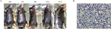

| 时间(d) | 对照组(15只) (mm3, | 黄芪多糖组(15只) (mm3, | t值 | P值 |
|---|---|---|---|---|
| 5 | 31.01±11.25 | 31.82±9.16 | 0.217 | 0.830 |
| 8 | 66.79±25.44 | 57.72±15.18 | 1.185 | 0.246 |
| 11 | 135.81±42.98 | 92.13±22.53 | 3.583 | 0.001 |
| 14 | 269.66±96.39 | 169.55±49.91 | 3.572 | 0.001 |
| 17 | 441.24±129.35 | 285.25±71.80 | 4.084 | <0.001 |
| 20 | 690.56±184.61 | 484.68±142.61 | 3.572 | 0.001 |
| 23 | 1139.76±323.36 | 756.18±129.28 | 4.266 | <0.001 |
| 26 | 1624.85±305.09 | 1199.75±206.12 | 4.522 | <0.001 |
| 29 | 2059.54±167.77 | 1738.34±493.74 | 3.118 | 0.005 |
| 观察时间 (d) | 对照组(15只) | 黄芪多糖组(15只) | χ2值 | P值 | ||
|---|---|---|---|---|---|---|
| 生存数量(只) | 生存率(%) | 生存数量(只) | 生存率(%) | |||
| 0 | 15 | 100.0 | 15 | 100.0 | ||
| 23 | 15 | 100.0 | 15 | 100.0 | 12.180 | <0.001 |
| 26 | 13 | 86.7 | 15 | 100.0 | ||
| 29 | 9 | 60.0 | 14 | 93.3 | ||

| 变量 | 对照组(10只) (%, | 黄芪多糖组(10只) (%, | t值 | P值 |
|---|---|---|---|---|
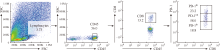
| CD8+/CD45+ | 16.92±3.29 | 16.84±2.87 | 0.059 | 0.954 |
| PD-1+CD8+/CD8+T | 83.41±1.95 | 78.81±2.61 | 4.467 | <0.001 |
| PD-1hiCD8+/CD8+ T | 31.16±5.84 | 21.52±1.94 | 4.597 | <0.001 |
| PD-1intCD8+/CD8+ T | 52.20±5.83 | 57.48±2.84 | 2.542 | 0.020 |
| 组别 | PD-1intCD8+T细胞(%, | PD-1hiCD8+T细胞(%, | ||||||
|---|---|---|---|---|---|---|---|---|
| MTG | Rhod-2 | Mito-SOX | TMRE | MTG | Rhod-2 | Mito-SOX | TMRE | |
| 对照组(10只) | 56.97±8.83 | 68.18±10.47 | 11.83±4.51 | 12.06±4.09 | 51.43±6.34 | 78.95±9.48 | 24.66±5.54 | 10.88±2.59 |
| 黄芪多糖组(10只) | 63.42±8.04 | 57.10±6.25 | 9.41±1.38 | 14.71±5.32 | 65.15±8.89 | 69.25±5.56 | 14.78±3.14 | 13.29±4.41 |
| t值 | 1.709 | 2.873 | 1.620 | 1.250 | 3.975 | 2.791 | 4.907 | 1.491 |
| P值 | 0.105 | 0.010 | 0.124 | 0.227 | <0.001 | 0.012 | <0.001 | 0.153 |
| 变量 | 对照组(10只) (%, | 黄芪多糖组(10只) (%, | t值 | P值 |
|---|---|---|---|---|
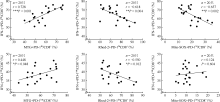
| INF-γ+/PD-1+CD8+ | 41.56±6.25 | 50.55±4.08 | 3.810 | 0.001 |
| INF-γ+/PD-1hiCD8+ | 58.87±6.54 | 70.48±3.62 | 4.910 | <0.001 |
| INF-γ+/PD-1intCD8+ | 36.97±3.30 | 40.76±4.32 | 2.204 | 0.041 |

| [1] | Siegel RL, Miller KD, Wagle NS, et al. Cancer statistics, 2023. CA Cancer J Clin, 2023, 73(1):17-48. doi:10.3322/caac.21763. |
| [2] | Han B, Zheng R, Zeng H, et al. Cancer incidence and mortality in China, 2022. J Natl Cancer Cent, 2024, 4(1):47-53. doi:10.1016/j.jncc.2024.01.006. |
| [3] |
Song P, Zhang J, Shang C, et al. Real-world evidenceand clinical observations of the treatment of advanced non-small cell lung cancer with PD-1/PD-L1 inhibitors. Sci Rep, 2019, 9(1):4278. doi:10.1038/s41598-019-40748-7.
pmid: 30862891 |
| [4] |
Okiyama N, Tanaka R. Immune-related adverse events in various organs caused by immune checkpoint inhibitors. Allergol Int, 2022, 71(2):169-178. doi:10.1016/j.alit.2022.01.001.
pmid: 35101349 |
| [5] | 陈佳骏, 邱磊, 王蕾, 等. 中药在治疗非小细胞肺癌中的潜在作用研究进展. 中成药, 2024, 46(1):204-210. doi:10.3969/j.issn.1001-1528.2024.01.035. |
| [6] | Kong FM, Chen TQ, Li XJ, et al. The Current Application and Future Prospects of Astragalus Polysaccharide Combined with Cancer Immunotherapy:A Review. Front Pharmacol, 2021, 12:737674. doi:10.3389/fphar.2021.737674. |
| [7] |
Franco F, Jaccard A, Romero P, et al. Metabolic and epigenetic regulation of T-cell exhaustion. Nat Metab, 2020, 2(10):1001-1012. doi:10.1038/s42255-020-00280-9.
pmid: 32958939 |
| [8] | Bamodu OA, Kuo KT, Wang CH, et al. Astragalus polysaccharides (PG2) Enhances the M1 Polarization of Macrophages, Functional Maturation of Dendritic Cells, and T Cell-Mediated Anticancer Immune Responses in Patients with Lung Cancer. Nutrients, 2019, 11(10): 2264. doi:10.3390/nu11102264. |
| [9] | Wang J, Zhang T, Cheng X. Regulatory Effect of Astragalus Polysacharin on Expresion of PD-1/PD-Ls Molecules in Melanoma Mice. Acta Universitatis Traditionis Medicalis Sinensis, 2014, 28 (5): 74-79. doi:10.16306/j.1008-861x.2014.05.019. |
| [10] | Gong Q, Yu H, Ding G, et al. Suppression of stemness and enhancement of chemosensibility in the resistant melanoma were induced by Astragalus polysaccharide through PD-L1 downregulation. Eur J Pharmacol, 2022, 916:174726. doi:10.1016/j.ejphar.2021.174726. |
| [11] | Zhang H, Huang H, Wu S, et al. Single-cell RNA sequencing reveals the effects of anti-PD-L 1 therapy on 3LL lung cancer model and its tumor microenvironment. Med Oncol, 2023, 40(10):285. doi:10.1007/s12032-023-02156-w. |
| [12] | Lv LL, Zhai JW, Wu JJ, et al. High CD38 expression defines a mitochondrial function-adapted CD8+ T cell subset with implications for lung cancer immunotherapy. Cancer Immunol Immunother, 2025, 74(2):49. doi:10.1007/s00262-024-03881-5. |
| [13] | Zhao LC, Zhong YT, Liang J, et al. Effect of Astragalus Polysaccharide on the Expression of VEGF and EGFR in Mice with Lewis Transplantable Lung Cancer. J Coll Physicians Surg Pak, 2019, 29(4): 392-394. doi:10.29271/jcpsp.2019.04.392. |
| [14] | Lim SM, Park HB, Jin JO. Polysaccharide from Astragalus membranaceus promotes the activation of human peripheral blood and mouse spleen dendritic cells. Chin J Nat Med, 2021, 19(1):56-62. doi:10.1016/S1875-5364(21)60006-7. |
| [15] | Liao CH, Yong CY, Lai GM, et al. Astragalus Polysaccharide (PG2) Suppresses Macrophage Migration Inhibitory Factor and Aggressiveness of Lung Adenocarcinoma Cells. Am J Chin Med, 2020, 48(6): 1491-1509. doi:10.1142/S0192415X20500731. |
| [16] |
Phacharapiyangkul N, Wu LH, Lee WY, et al. The extracts of Astragalus membranaceus enhance chemosensitivity and reduce tumor indoleamine 2, 3-dioxygenase expression. Int J Med Sci, 2019, 16(8): 1107-1115. doi:10.7150/ijms.33106.
pmid: 31523173 |
| [17] | Chang FL, Tsai KC, Lin TY, et al. Astragalus membranaceus-Derived Anti-Programmed Death-1 Monoclonal Antibodies with Immunomodulatory Therapeutic Effects against Tumors. Biomed Res Int, 2020, 2020:3415471. doi:10.1155/2020/3415471. |
| [18] |
Zhang Y, Wang K, Qin C, et al. Mitochondria dysfunction in CD8+ T cells as an important contributing factor for cancer development and a potential target for cancer treatment: a review. J Exp Clin Cancer Res, 2022, 41(1):227. doi:10.1186/s13046-022-02439-6.
pmid: 35864520 |
| [19] |
Hashimoto M, Kamphorst AO, Im SJ, et al. CD8 T Cell Exhaustion in Chronic Infection and Cancer: Opportunities for Interventions. Annu Rev Med, 2018, 69:301-318. doi:10.1146/annurev-med-012017-043208.
pmid: 29414259 |
| [20] | Zehn D, Thimme R, Lugli E, et al. ‘Stem-like’ precursors are the fount to sustain persistent CD8+ T cell responses. Nat Immunol, 2022, 23(6):836-847. doi:10.1038/s41590-022-01219-w. |
| [21] |
Scharping NE, Rivadeneira DB, Menk AV, et al. Mitochondrial stress induced by continuous stimulation under hypoxia rapidly drives T cell exhaustion. Nat Immunol, 2021, 22(2): 205-215. doi:10.1038/s41590-020-00834-9.
pmid: 33398183 |
| [22] | Yu YR, Imrichova H, Wang H, et al. Disturbed mitochondrial dynamics in CD8+ TILs reinforce T cell exhaustion. Nat Immunol, 2020, 21(12):1540-1551. doi:10.1038/s41590-020-0793-3. |
| [23] |
Soto-Heredero G, Desdín-Micó G, Mittelbrunn M. Mitochondrial dysfunction defines T cell exhaustion. Cell Metab, 2021, 33(3):470-472. doi:10.1016/j.cmet.2021.02.010.
pmid: 33657392 |
| [24] | Kumar A, Chamoto K, Chowdhury PS, et al. Tumors attenua-ting the mitochondrial activity in T cells escape from PD-1 blockade therapy. Elife, 2020, 9:e52330. doi:10.7554/eLife.52330. |
| [25] | Ma Q, Xu Y, Tang L, et al. Astragalus Polysaccharide Attenuates Cisplatin-induced Acute Kidney Injury by Suppressing Oxidative Damage and Mitochondrial Dysfunction. Biomed Res Int, 2020, 2020:2851349. doi:10.1155/2020/2851349. |
| [26] | Thibaut R, Bost P, Milo I, et al. Bystander IFN-γ activity promotes widespread and sustained cytokine signaling altering the tumor microenvironment. Nat Cancer, 2020, 1(3):302-314. doi:10.1038/s43018-020-0038-2. |
| [27] | Hoekstra ME, Bornes L, Dijkgraaf FE, et al. Long-distance modulation of bystander tumor cells by CD8+ T cell-secreted IFNγ. Nat Cancer, 2020, 1(3):291-301. doi:10.1038/s43018-020-0036-4. |
| [1] | Multidisciplinary Diagnosis and Treatment Branch of Chinese Antituberculosis Association , National Clinical Research Center for Infectious Disease/Shenzhen Third People’s Hospital, Beijing Chao-Yang Hospital , Capital Medical University , Guangdong Lung Cancer Institute . Expert consensus on the diagnosis and treatment of coexistent pulmonary tuberculosis and lung cancer [J]. Journal of Tuberculosis and Lung Disease, 2025, 6(5): 495-515. |
| [2] | Wang Ning, Wang Baohua. The burden, prevention and control status, and prospects of global lung cancer [J]. Journal of Tuberculosis and Lung Disease, 2025, 6(3): 256-260. |
| [3] | Chen Jing, Qin Yali, Wang Mingdong, Yang Rubin, Wang Qian, Peng Yanqing, Qiu Jiyao, Zhang Xiao, Zhou Xinai. The value of QuantiFERON-TB Gold Plus in the clinical diagnosis of active pulmonary tuberculosis [J]. Journal of Tuberculosis and Lung Disease, 2025, 6(1): 61-67. |
| [4] | Zhu Yixing, Chang De. Current status and prospects of management of chronic respiratory diseases [J]. Journal of Tuberculosis and Lung Disease, 2024, 5(6): 567-572. |
| [5] | Chen Xinxin, Liu Guofeng, Yang Yingzi, Hu Yachen, Jin Ying, Li Yumei. Correlation analysis of physical activity levels and symptom clusters in lung cancer chemotherapy patients during hospitalization [J]. Journal of Tuberculosis and Lung Disease, 2024, 5(3): 197-206. |
| [6] | Pan Qiong, Tian Yayuan, Tong Fei. Effect of early limb active exercise on incidence of pulmonary embolism among patients received lung cancer surgery [J]. Journal of Tuberculosis and Lung Disease, 2024, 5(3): 212-218. |
| [7] | Zi Xiaohui, Wu Peng, Zheng Sufei, Sun Nan, He Jie. Meta-analysis of the impact of low-dose CT screening on population lung cancer-specific mortality and all-cause mortality [J]. Journal of Tuberculosis and Lung Disease, 2024, 5(2): 106-112. |
| [8] | Tang Ke, Yuan Xiaodong, Zhang Laixing. Clinical education assisted by three-dimensional model for detecting pulmonary ground glass nodule [J]. Journal of Tuberculosis and Lung Disease, 2023, 4(5): 346-351. |
| [9] | Fan Panyu, Li Yumei, Yang Lei, Fan Jiaxin. Study on influencing factors of sleep quality in elderly patients with advanced lung cancer undergoing chemotherapy [J]. Journal of Tuberculosis and Lung Disease, 2023, 4(3): 210-215. |
| [10] | LI Xiang-nan, ZHANG Xiao-ju. Research progress of tumor ablation in lung cancer [J]. Journal of Tuberculosis and Lung Disease, 2021, 2(3): 256-261. |
| [11] | ZHANG Qun-cheng, LI Xiang-nan, XU Zhi-wei, SUN Guan-nan, YANG Hui-zhen, CHENG Dong-jun, ZHANG Xiao-ju. Diagnostic value of bronchoscopic cryobiopsy for diagnosing peripheral lung nodule [J]. Journal of Tuberculosis and Lung Disease, 2021, 2(3): 205-209. |
| [12] | LIU Hai-yang, ZHANG Xiao-ju. Application of predictive model in early diagnosis of lung cancer [J]. Journal of Tuberculosis and Lung Disease, 2021, 2(3): 262-266. |
| [13] | YANG Hui-zhen, ZHANG Qun-cheng, ZHANG Xiao-ju. Advances in clinical application of endobronchial ultrasound-guided transbronchial needle aspiration [J]. Journal of Tuberculosis and Lung Disease, 2021, 2(3): 267-272. |
| [14] | CUI Kai, ZHANG Xiao-ju. Research progress on the application of biomarkers in differential diagnosis of benign and malignant pulmonary nodules [J]. Journal of Tuberculosis and Lung Disease, 2021, 2(3): 273-276. |
| [15] | XIN Wu-qun, CHEN Xiao, TANG Jin-xing, XU Gao-jun, ZHOU Zhen-qiang, HE Yi. Factors of persistent cough secondary to thoracoscopic resection of lung cancer [J]. Journal of Tuberculosis and Lung Disease, 2021, 2(1): 31-37. |
| Viewed | ||||||
|
Full text |
|
|||||
|
Abstract |
|
|||||
 京公安网备11010202008787号
Total visitors: Visitors of today: Now online:
京公安网备11010202008787号
Total visitors: Visitors of today: Now online:
 This work is licensed under Creative Commons Attribution 3.0 License.