
结核与肺部疾病杂志 ›› 2024, Vol. 5 ›› Issue (5): 388-397.doi: 10.19983/j.issn.2096-8493.2024084
云南省传染性疾病临床医学中心, 云南省医院协会呼吸内科专业委员会, 昆明市医学会肺结节早诊早治专业委员会
收稿日期:2024-05-09
出版日期:2024-10-20
发布日期:2024-10-14
通信作者:
陆霓虹,Email:基金资助:Yunnan Provincial Clinical Medical Center for Infectious Disease, Professional Committee of Respiratory Medicine of Yunnan Hospital Association, Professional Committee of Early Diagnosis and Treatment of Pulmonary Nodules of Kunming Medical Association
Received:2024-05-09
Online:2024-10-20
Published:2024-10-14
Contact:
Lu Nihong,Email:Supported by:摘要:
肺结节筛查计划推广及低剂量胸部CT日益普及,让更多肺结节患者得到早期诊断。但如何精准识别高危肺结节,让患者得到准确诊断和规范化诊疗,是目前肺结节诊疗的热点和难点。2015年首部“肺结节诊治专家共识”发表后,各个地区结合当地实情相继完善了肺结节诊治与管理策略,参考国内外文献成果及现有的肺结节诊疗专家共识,结合筛查肺结节的临床诊疗经验,经过呼吸、感染、影像、病理等领域专家的多轮会议研讨,最终制定了这部肺结节诊治专家共识。通过阐述肺结节的诊治要点、治疗方法选择,旨在让呼吸学科及相关学科医师对肺结节规范诊疗有一个全面认识,也为各级医院开展肺结节规范诊治工作提供参考依据。
中图分类号:
云南省传染性疾病临床医学中心, 云南省医院协会呼吸内科专业委员会, 昆明市医学会肺结节早诊早治专业委员会. 肺结节规范化诊治专家共识[J]. 结核与肺部疾病杂志, 2024, 5(5): 388-397. doi: 10.19983/j.issn.2096-8493.2024084
Yunnan Provincial Clinical Medical Center for Infectious Disease, Professional Committee of Respiratory Medicine of Yunnan Hospital Association, Professional Committee of Early Diagnosis and Treatment of Pulmonary Nodules of Kunming Medical Association. Expert consensus on standardized diagnosis and treatment of pulmonary nodules[J]. Journal of Tuberculosis and Lung Disease, 2024, 5(5): 388-397. doi: 10.19983/j.issn.2096-8493.2024084
表1
良、恶性肺结节外观及内部特征对比
| 项目 | 良性结节 | 恶性结节 |
|---|---|---|
| 形态特征 | 圆形、类圆形、三角形 | 圆形、类圆形 |
| 边缘特征 | 光滑、平整,长毛刺 | 分叶、短毛刺、胸膜牵拉、胸膜附着、胸膜凹陷、胸膜尾征 |
| 交界面特征 | 清楚、整齐,甚至光整 | 清楚但毛糙 |
| 内部特征 | 呈脂肪密度、钙化、低密度液化 | 血管征(扭曲/扩张/僵硬)、空泡征及囊腔型 |
| 邻近结构 | 周围血管分布走向正常或绕行 | 胸膜凹陷征、血管集束征 |
| 血供(强化程度) | 无强化或轻度强化,硬化性肺泡细胞瘤、活动性炎性结节及血管性病变可呈明显强化,强化峰值多出现在动脉期 | 中度以上均匀或不均匀强化,强化峰值多出现在延迟期 |

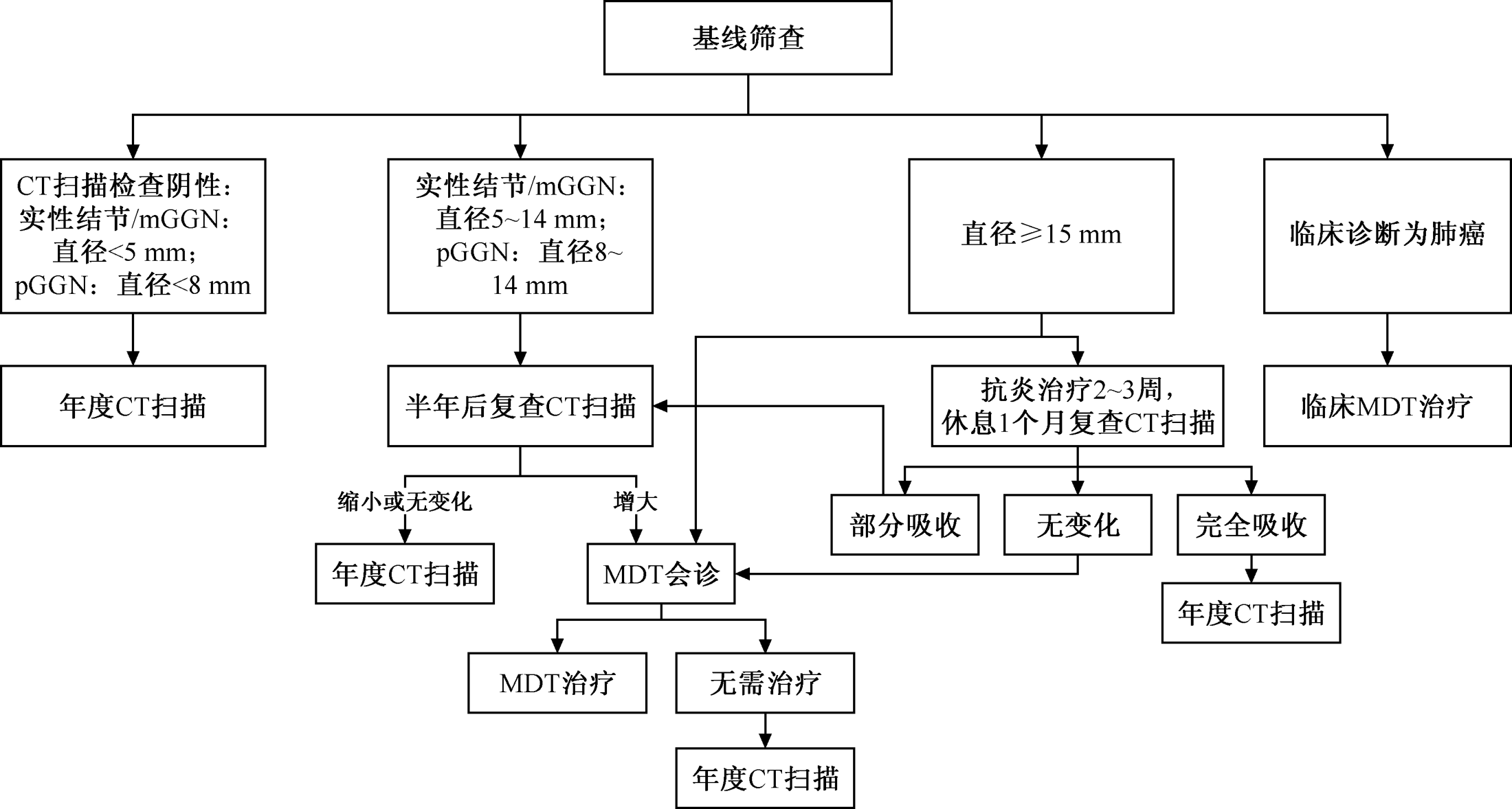
图1
基线筛查发现的肺结节随访策略 注 mGGN:部分实性结节; pGGN:非实性结节; CT:计算机断层扫描; MDT:多学科诊疗
| [1] |
Mazzone PJ, Lam L. Evaluating the Patient With a Pulmonary Nodule: A Review. JAMA, 2022, 327(3): 264-273. doi:10.1001/jama.2021.24287.
pmid: 35040882 |
| [2] | 周清华, 范亚光, 王颖, 等. 中国肺部结节分类、诊断与治疗指南(2016年版). 中国肺癌杂志, 2016, 19(12): 793-798. doi:10.3779/j.issn.1009-3419.2016.12.12. |
| [3] |
Walter K. Pulmonary Nodules. JAMA, 2021, 326(15): 1544. doi:10.1001/jama.2021.12319.
pmid: 34665202 |
| [4] | 中国肺癌早诊早治专家组, 中国西部肺癌研究协作中心. 中国肺癌低剂量CT筛查指南(2023年版). 中国肺癌杂志, 2023, 26(1): 1-9. doi:10.3779/j.issn.1009-3419.2023.102.10. |
| [5] | 陈亚男, 陈武飞, 滑炎卿. 肺结节倍增时间的CT研究进展. 中华解剖与临床杂志, 2017, 22(6): 522-527. doi:10.3760/cma.j.issn.2095-7041.2017.06.016. |
| [6] | 孟祥鹿, 幸子健, 卢山. 基于深度学习的肺结节分类分割算法及其在不同CT重建算法下的效能评估. 中华医学杂志, 2021, 101(7): 476-480. doi:10.3760/cma.j.cn112137-20201123-03171. |
| [7] | 刘敬伟, 张西宁. 肺结节诊断方法的最新进展. 中华外科杂志, 2022, 60(5): 498-503. doi:10.3760/cma.j.cn112139-20211123-00551. |
| [8] | Zhou SC, Wang YJ, Ai T, et al. Diagnosis of solitary pulmonary lesions with intravoxel incoherent motion diffusion-weighted MRI and semi-quantitative dynamic contrast-enhanced MRI. Clin Radiol, 2019, 74(5): 409.e7-e16. doi:10.1016/j.crad.2018.12.022. |
| [9] | 周舒畅, 王玉锦, 黄璐, 等. 扩散峰度成像与扩散加权成像在难鉴别孤立性肺结节良恶性判定价值的比较研究. 中华放射学杂志, 2019, 53(3): 200-204. doi:3760/cma.j.issn.1005-1201.2019.03.008. |
| [10] | Gould MK, Donington J, Lynch WR, et al. Evaluation of individuals with pulmonary nodules: when is it lung cancer? Diagnosis and management of lung cancer, 3rd ed: American College of Chest Physicians evidence-based clinical practice guidelines. Chest, 2013, 143(5 Suppl): e93S-e120S. doi:10.1378/chest.12-2351. |
| [11] |
Volpi S, Ali J M, Tasker A, et al. The role of positron emission tomography in the diagnosis, staging and response assessment of non-small cell lung cancer. Ann Transl Med, 2018, 6(5): 95. doi:10.21037/atm.2018.01.25.
pmid: 29666818 |
| [12] |
Chun EJ, Lee HJ, Kang WJ, et al. Differentiation between malignancy and inflammation in pulmonary ground-glass nodules: The feasibility of integrated (18)F-FDG PET/CT. Lung Cancer, 2009, 65(2): 180-186. doi:10.1016/j.lungcan.2008.11.015.
pmid: 19155090 |
| [13] |
Heyneman LE, Patz EF. PET imaging in patients with bronchioloalveolar cell carcinoma. Lung Cancer, 2002, 38(3): 261-266. doi:10.1016/s0169-5002(02)00221-0.
pmid: 12445747 |
| [14] | 吴少虹, 郭莉莉. 影像组学在肺癌中的应用. 放射学实践, 2023, 38(6): 778-782. doi:10.13609/j.cnki.1000-0313.2023.06.019. |
| [15] | Yang DW, Zhang Y, Hong QY, et al. Role of a serum-based biomarker panel in the early diagnosis of lung cancer for a cohort of high-risk patients. Cancer, 2015, 121 Suppl 17:3113-3121. doi:10.1002/cncr.29551. |
| [16] | 孙硕, 王锋, 何立, 等. 液体活检生物标志物及其联合影像学在肺癌早期诊断中应用的研究进展. 中国胸心血管外科临床杂志, 2023, 30(2): 313-319. doi:10.7507/1007-4848.202205044. |
| [17] | 刘宝东, 陈海泉, 刘伦旭, 等. 肺结节多学科微创诊疗中国专家共识. 中国胸心血管外科临床杂志, 2023, 30(8):1061-1074. doi:10.7507/1007-4848.202306006. |
| [18] | Han Y, Kim HJ, Kong KA, et al. Diagnosis of small pulmonary lesions by transbronchial lung biopsy with radial endobronchial ultrasound and virtual bronchoscopic navigation versus CT-guided transthoracic needle biopsy: A systematic review and meta-analysis. PLoS One, 2018, 13(1): e0191590. doi:10.1371/journal.pone.0191590. |
| [19] |
Wang Memoli JS, Nietert PJ, Silvestri GA. Meta-analysis of guided bronchoscopy for the evaluation of the pulmonary nodule. Chest, 2012, 142(2): 385-393. doi:10.1378/chest.11-1764.
pmid: 21980059 |
| [20] |
Folch EE, Pritchett MA, Nead MA, et al. Electromagnetic Navigation Bronchoscopy for Peripheral Pulmonary Lesions: One-Year Results of the Prospective, Multicenter NAVIGATE Study. J Thorac Oncol, 2019, 14(3): 445-458. doi:10.1016/j.jtho.2018.11.013.
pmid: 30476574 |
| [21] | Deng CJ, Dai FQ, Qian K, et al. Clinical updates of approaches for biopsy of pulmonary lesions based on systematic review. BMC Pulm Med, 2018, 18(1):146. doi:10.1186/s12890-018-0713-6. |
| [22] | 刘冰, 林钢, 刘敬伟, 等. 2cm单孔胸腔镜肺叶切除术的临床应用. 中国胸心血管外科临床杂志, 2017, 24(7): 566-568. doi:10.7507/1007-4848.201610035. |
| [23] |
Gonzalez-Rivas D, Paradela M, Fernandez R, et al. Uniportal video-assisted thoracoscopic lobectomy: two years of experience. Ann Thorac Surg, 2013, 95(2): 426-32. doi:10.1016/j.athoracsur.2012.10.070.
pmid: 23219257 |
| [24] |
Wahidi MM, Govert JA, Goudar RK, et al. Evidence for the treatment of patients with pulmonary nodules: when is it lung cancer?: ACCP evidence-based clinical practice guidelines (2nd edition). Chest, 2007, 132(3 Suppl): 94s-107s. doi:10.1378/chest.07-1352.
pmid: 17873163 |
| [25] |
Lee GD, Park CH, Park HS, et al. Lung Adenocarcinoma Invasiveness Risk in Pure Ground-Glass Opacity Lung Nodules Smaller than 2 cm. Thorac Cardiovasc Surg, 2019, 67(4): 321-328. doi:10.1055/s-0037-1612615.
pmid: 29359309 |
| [26] | 范丽, 望云, 周秀秀, 等. 孤立性肺结节的影像诊断思路及处理策略. 中华放射学杂志, 2023, 57(2): 235-238. doi:10.3760/cma.j.cn112149-20220708-00590. |
| [27] | Gao F, Sun Y, Zhang G, et al. CT characterization of different pathological types of subcentimeter pulmonary ground-glass nodular lesions. Br J Radiol, 2019, 92(1094): 20180204. doi:10.1259/bjr.20180204. |
| [28] | Liang J, Xu XQ, Xu H, et al. Using the CT features to differentiate invasive pulmonary adenocarcinoma from pre-invasive lesion appearing as pure or mixed ground-glass nodules. Br J Radiol, 2015, 88(1053): 20140811. doi:10.1259/bjr.20140811. |
| [29] |
Mets OM, De Jong PA, Scholten ET, et al. Subsolid pulmonary nodule morphology and associated patient characteristics in a routine clinical population. Eur Radiol, 2017, 27(2): 689-696. doi:10.1007/s00330-016-4429-9.
pmid: 27255399 |
| [30] |
Silva M, Sverzellati N, Manna C, et al. Long-term surveillance of ground-glass nodules: evidence from the MILD trial. J Thorac Oncol, 2012, 7(10): 1541-1546. doi:10.1097/JTO.0b013e3182641bba.
pmid: 22968185 |
| [31] |
Wu F, Tian SP, Jin X, et al. CT and histopathologic characteristics of lung adenocarcinoma with pure ground-glass nodules 10 mm or less in diameter. Eur Radiol, 2017, 27(10): 4037-4043. doi:10.1007/s00330-017-4829-5.
pmid: 28386719 |
| [32] | Xiang W, Xing Y, Jiang S, et al. Morphological factors differen-tiating between early lung adenocarcinomas appearing as pure ground-glass nodules measuring ≤10 mm on thin-section computed tomography. Cancer Imaging, 2014, 14(1): 33. doi:10.1186/s40644-014-0033-x. |
| [33] | Tammemagi MC, Schmidt H, Martel S, et al. Participant selection for lung cancer screening by risk modelling (the Pan-Canadian Early Detection of Lung Cancer [PanCan] study): a single-arm, prospective study. Lancet Oncol, 2017, 18(11): 1523-1531. doi:10.1016/S1470- 2045(17)30597-1. |
| [34] |
Macmahon H, Naidich DP, Goo JM, et al. Guidelines for Management of Incidental Pulmonary Nodules Detected on CT Images: From the Fleischner Society 2017. Radiology, 2017, 284(1): 228-243. doi:10.1148/radiol.2017161659.
pmid: 28240562 |
| [35] | Wood DE, Kazerooni EA, Aberle D, et al. NCCN Guidelines Insights: Lung Cancer Screening, Version 1.2022. J Natl Compr Canc Netw, 2022, 20(7): 754-764. doi:10.6004/jnccn.2022.0036. |
| [36] | Haiman CA, Stram DO, Wilkens LR, et al. Ethnic and racial differences in the smoking-related risk of lung cancer. N Engl J Med, 2006, 354(4): 333-342. doi:10.1056/NEJMoa033250. |
| [37] | 吕章艳, 谭锋维, 林春青, 等. 肺癌风险预测模型构建与验证的系统综述. 中华预防医学杂志, 2020, 54(4): 430-437. doi:10.3760/cma.j.cn112150-20190523-00415. |
| [38] |
Swensen SJ, Silverstein MD, Ilstrup DM, et al. The probability of malignancy in solitary pulmonary nodules. Application to small radiologically indeterminate nodules. Arch Intern Med, 1997, 157(8): 849-855.
pmid: 9129544 |
| [39] |
Gould MK, Ananth L, BarnetT PG. A clinical model to estimate the pretest probability of lung cancer in patients with solitary pulmonary nodules. Chest, 2007, 131(2): 383-388. oi: 10.1378/chest. 06-1261.
doi: 10.1378/chest.06-1261 pmid: 17296637 |
| [40] | 李运, 陈克终, 隋锡朝, 等. 孤立性肺结节良恶性判断数学预测模型的建立. 北京大学学报(医学版), 2011, 43(3): 450-454. doi:10.3969/j.issn.1671-167X.2011.03.027. |
| [41] | Mcwilliams A, Tammemagi MC, Mayo JR, et al. Probability of cancer in pulmonary nodules detected on first screening CT. N Engl J Med, 2013, 369(10): 910-919. doi:10.1056/NEJMoa1214726. |
| [42] | 肺结节诊治西北地区专家共识专家组, 陕西省抗癌协会微创治疗专业委员会, 陕西省老年学和老年医学学会肺癌专业委员会. 肺结节诊治西北地区专家共识(2021年版). 中国医药科学, 2021, 11(23): 16-22. doi:10.3969/j.issn.2095-0616.2021.23.007. |
| [43] |
Zhang Y, Deng C, Zheng Q, et al. Selective Mediastinal Lymph Node Dissection Strategy for Clinical T1N0 Invasive Lung Cancer: A Prospective, Multicenter, Clinical Trial. J Thorac Oncol, 2023, 18(7): 931-939. doi:10.1016/j.jtho.2023.02.010.
pmid: 36841542 |
| [44] | 中华医学会肿瘤学分会, 中华医学会杂志社. 中华医学会肺癌临床诊疗指南(2023版). 中华肿瘤杂志, 2023, 45(7): 539-574. doi:10.3760/cma.j.cn112152-20230510-00200. |
| [45] | Miyazaki T, Yamazaki T, Sato S, et al. Surgery or stereotactic body radiotherapy for metachronous primary lung cancer? A propensity score matching analysis. Gen Thorac Cardiovasc Surg, 2020, 68(11): 1305-1311. doi:10.1007/s11748-020-01394-3. |
| [46] |
Zhao L, Liu C, Xie G, et al. Multiple Primary Lung Cancers: A New Challenge in the Era of Precision Medicine. Cancer Manag Res, 2020, 12: 10361-10374. doi:10.2147/CMAR.S268081.
pmid: 33116891 |
| [47] | 郑加生, 叶欣. 中国肿瘤消融治疗的现状与未来. 中华医学杂志, 2017, 97(31): 2401-2403. doi:10.3760/cma.j.issn.0376-2491.2017.31.001. |
| [48] |
Koizumi T, Tsushima K, Tanabe T, et al. Bronchoscopy-Guided Cooled Radiofrequency Ablation as a Novel Intervention Therapy for Peripheral Lung Cancer. Respiration, 2015, 90(1): 47-55. doi:10.1159/000430825.
pmid: 26044954 |
| [49] |
Xie F, Zheng X, Xiao B, et al. Navigation Bronchoscopy-Guided Radiofrequency Ablation for Nonsurgical Peripheral Pulmonary Tumors. Respiration, 2017, 94(3): 293-298. doi:10.1159/000477764.
pmid: 28683443 |
| [50] | Genshaft SJ, Suh RD, Abtin F, et al. Society of Interventional Radiology Quality Improvement Standards on Percutaneous Ablation of Non-Small Cell Lung Cancer and Metastatic Disease to the Lungs. J Vasc Interv Radiol, 2021, 32(8): 1242. e1-1242.e10. doi:10.1016/j.jvir.2021.04.027. |
| [51] | 中国临床肿瘤学会CSCO肿瘤消融治疗专家委员会, 中国医师协会肿瘤消融治疗技术专家组, 中国抗癌协会肿瘤消融治疗专业委员会, 等. 影像引导下热消融治疗原发性和转移性肺部肿瘤临床实践指南(2021年版). 中华内科杂志, 2021, 60(12): 1088-1105. doi:10.3760/cma.j.cn112138-20210814-00554. |
| [52] |
Hasegawa T, Kondo C, Sato Y, et al. Pathologic Diagnosis and Genetic Analysis of a Lung Tumor Needle Biopsy Specimen Obtained Immediately After Radiofrequency Ablation. Cardiovasc Intervent Radiol, 2018, 41(4): 594-602. doi:10.1007/s00270-017-1845-4.
pmid: 29164309 |
| [53] |
Wang J, Ni Y, Yang X, et al. Diagnostic ability of percutaneous core biopsy immediately after microwave ablation for lung ground-glass opacity. J Cancer Res Ther, 2019, 15(4): 755-759. doi:10.4103/jcrt.JCRT_399_19.
pmid: 31436228 |
| [54] |
Chi J, Ding M, Wang Z, et al. Pathologic Diagnosis and Genetic Analysis of Sequential Biopsy Following Coaxial Low-Power Microwave Thermal Coagulation For Pulmonary Ground-Glass Opacity Nodules. Cardiovasc Intervent Radiol, 2021, 44(8): 1204-1213. doi:10.1007/s00270-021-02782-9.
pmid: 33825064 |
| [1] | 阎庆虎, 薛峰, 于泳, 秦毅, 阎庆梅, 崔嘉. 超声引导下微波消融技术在局限性结核病变治疗中的价值分析[J]. 结核与肺部疾病杂志, 2025, 6(1): 55-60. |
| [2] | 陈静, 秦娅莉, 王明栋, 杨儒斌, 王倩, 彭燕清, 邱继瑶, 张晓, 周昕艾. QuantiFERON-TB Gold Plus检测活动性肺结核的效能分析[J]. 结核与肺部疾病杂志, 2025, 6(1): 61-67. |
| [3] | 顾金花, 张盼盼. 三种结核分枝杆菌检测方法在某综合医院的应用价值评估[J]. 结核与肺部疾病杂志, 2025, 6(1): 68-72. |
| [4] | 廖影, 庞艳, 赵静, 何高琴, 游茂林, 王蕾. 2018—2023年重庆市梁平区肺结核患者报告情况及发现延迟特征分析[J]. 结核与肺部疾病杂志, 2025, 6(1): 8-13. |
| [5] | 张莹, 郭春辉. 结核性气管支气管狭窄的治疗研究进展[J]. 结核与肺部疾病杂志, 2025, 6(1): 87-93. |
| [6] | 欧庆芬. 非结核分枝杆菌肺病的CT诊断及鉴别诊断[J]. 结核与肺部疾病杂志, 2024, 5(S): 13-14. |
| [7] | 厉银锋, 叶雅利, 杨禧龙, 张海啸. 电针治疗对长期过量使用唑吡坦引起呼吸抑制的干预策略[J]. 结核与肺部疾病杂志, 2024, 5(S): 52-55. |
| [8] | 赵静, 郑一兵. 肺炎支原体肺炎的影像学表现及CT鉴别诊断效果[J]. 结核与肺部疾病杂志, 2024, 5(S): 73-75. |
| [9] | 秦海松. 多排螺旋CT在早期肺癌诊断中的敏感度观察[J]. 结核与肺部疾病杂志, 2024, 5(S): 76-78. |
| [10] | 刘园, 刘丽, 冯立君, 王小娜. 基于PCR方法诊断血清中布鲁氏菌在临床实验室应用价值的研究[J]. 结核与肺部疾病杂志, 2024, 5(S): 89-92. |
| [11] | 夏莎莎. 急诊护理干预在慢性阻塞性肺疾病患者中的急诊治疗效果[J]. 结核与肺部疾病杂志, 2024, 5(S): 108-110. |
| [12] | 彭莉. 慢性肾衰竭合并肺部感染患者治疗进展[J]. 结核与肺部疾病杂志, 2024, 5(S): 165-168. |
| [13] | 马晓玲, 赵永年, 段丽丽, 刘新文. 新疆生产建设兵团2016—2022年肺结核患者不良转归列线图预测模型的构建与验证[J]. 结核与肺部疾病杂志, 2024, 5(6): 552-559. |
| [14] | 孙慧娟, 苏伟, 陈伟. 利福平耐药结核病患者不良治疗结局及其影响因素研究进展[J]. 结核与肺部疾病杂志, 2024, 5(6): 573-582. |
| [15] | 曾琴, 曾凡清, 何坤, 杨红红, 刘敏. 常规病理学镜检、抗酸染色镜检及TB-DNA在浅表淋巴结结核诊断中的价值[J]. 结核与肺部疾病杂志, 2024, 5(5): 461-467. |
| 阅读次数 | ||||||
|
全文 |
|
|||||
|
摘要 |
|
|||||
 京公安网备11010202008787号
ip访问总数: ip当日访问总数: 当前在线人数:
京公安网备11010202008787号
ip访问总数: ip当日访问总数: 当前在线人数:
 本作品遵循Creative Commons Attribution 3.0 License授权许可