结核与肺部疾病杂志 ›› 2025, Vol. 6 ›› Issue (4): 432-435.doi: 10.19983/j.issn.2096-8493.20250043

• 研究论文 • 上一篇    下一篇

高海拔环境下肺结核与细菌性肺炎的超声影像学特征比较

柳静, 赵月娟()   

  1. 云南省传染病医院医学影像科,昆明 650301
  • 收稿日期:2025-03-10 出版日期:2025-08-20 发布日期:2025-08-08
  • 通信作者: 赵月娟,Email:13888378206@163.com
  • 基金资助:
    云南省科技厅科技重大专项计划(202102AA310005-20)

Comparison of ultrasound imaging characteristics between pulmonary tuberculosis and bacterial pneumonia in high-altitude environments

Liu Jing, Zhao Yuejuan()   

  1. Department of Medical Imaging, Infectious Disease Hospital of Yunnan Province, Kunming 650301, China
  • Received:2025-03-10 Online:2025-08-20 Published:2025-08-08
  • Contact: Zhao Yuejuan, Email: 13888378206@163.com
  • Supported by:
    Yunnan Provincial Department of Science and Technology Major Science and Technology Special Projects Program(202102AA310005-20)

摘要:

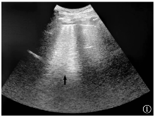
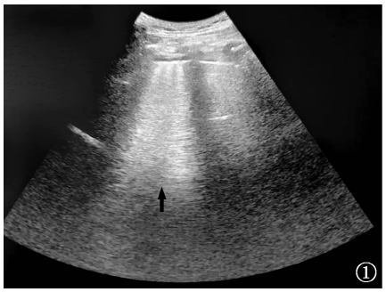
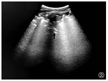
目的: 探讨高海拔环境下胸膜下肺结核与细菌性肺炎的超声影像学特征。方法: 采用回顾性研究方法,选取2024年1—12月云南省传染病医院确诊的47例胸膜下肺结核患者(肺结核组)及40例细菌性肺炎患者(肺炎组)作为研究对象。所有患者均接受常规超声检查,比较两组病灶形态(楔形、类圆形、不规则形)、碎片征(局部回声不均伴散在强光点)、瀑布征(层状无回声区与胸膜线平行延伸,记录其延伸范围及层状结构的清晰度)、液化坏死灶(无回声区内部可见气体强回声或“彗尾征”)等超声影像学特征。结果: 肺结核组中,楔形病灶占61.7%(29/47),类圆形病灶占29.8%(14/47),不规则形病灶占8.5%(4/47);肺炎组中,楔形病灶占62.5%(25/40),类圆形病灶占37.5%(15/40),两组在病灶形态分布上比较,差异无统计学意义(Fisher精确概率法,P>0.05)。肺结核组出现碎片征者占29.8%(14/47),明显低于肺炎组的82.5%(33/40),差异有统计学意义(χ2=14.490,P=0.001)。肺结核组出现“瀑布征”者占61.7%(29/47),明显高于肺炎组的27.5%(11/40),差异有统计学意义(χ2=10.177,P=0.001)。肺结核组出现液化坏死灶者占80.9%(38/47),明显高于肺炎组的25.0%(10/40),差异有统计学意义(χ2=28.301,P=0.001)。肺结核组有周边血流信号者占68.1%(32/47),有中央型血流者0例;肺炎组有周边血流信号者占77.5%(31/40),有中央型血流者占2.5%(1/40),差异无统计学意义(χ2=0.976,P=0.317)。结论: 高海拔环境下肺结核超声影像以层状无回声(瀑布征)伴气体强回声为特征,而细菌性肺炎以碎片征及小片液化灶为主,血流信号均以周边分布为主。

关键词: 结核,肺, 肺炎,细菌性, 超声检查, 疾病特征, 对比研究

Abstract:

Objective: To investigate the ultrasound imaging features of subpleural pulmonary tuberculosis and bacterial pneumonia in a high-altitude environment. Methods: Using a retrospective research method, 47 patients diagnosed with subpleural pulmonary tuberculosis (pulmonary tuberculosis group) and 40 patients diagnosed with bacterial pneumonia (pneumonia group) at Yunnan Provincial Infectious Disease Hospital from January to December 2024 were selected as the study subjects. All patients underwent routine ultrasound examination to compare the ultrasound imaging features of the two groups, including lesion morphology (wedge-shaped, quasi circular, irregular), fragmentation sign (uneven local echoes with scattered bright spots), waterfall sign (layered hypoechoic area extending parallel to the pleural line, recording its extension range and clarity of layered structure), and liquefaction necrosis lesion (gas hyperechoic or “comet tail sign” visible inside the hypoechoic area). Results: In the pulmonary tuberculosis group, wedge-shaped lesions accounted for 61.7% (29/47), round lesions for 29.8% (14/47), and irregular lesions for 8.5% (4/47). In the pneumonia group, wedge-shaped lesions accounted for 62.5% (25/40), and round lesions for 37.5% (15/40). There was no statistically significant difference in lesion morphology between the two groups (Fisher’s precision probability test, P>0.05). The proportion of patients with fragmented signs in the pulmonary tuberculosis group was 29.8% (14/47), significantly lower than the 82.5% (33/40) in the pneumonia group, and the difference was statistically significant (χ2=14.490, P=0.001). The incidence of “waterfall sign” in the pulmonary tuberculosis group was 61.7% (29/47), significantly higher than the 27.5% (11/40) in the pneumonia group, and the difference was statistically significant (χ2=10.177, P=0.001). The incidence of liquefied necrosis lesions in the pulmonary tuberculosis group was 80.9% (38/47), significantly higher than the 25.0% (10/40) in the pneumonia group, and the difference was statistically significant (χ2=28.301, P=0.001). 68.1% (32/47) of the pulmonary tuberculosis group had peripheral blood flow signals, and there were no cases of central blood flow; in the pneumonia group, 77.5% (31/40) had peripheral blood flow signals, while 2.5% (1/40) had central blood flow signals, with no statistically significant difference (χ2=0.976, P=0.317). Conclusion: The ultrasound imaging of pulmonary tuberculosis in high-altitude environments is characterized by layered hypoechoic (waterfall sign) with strong gas echoes, while bacterial pneumonia is mainly characterized by fragmented signs and small liquefied lesions, with blood flow signals mainly distributed in the periphery.

Key words: Tuberculosis, pulmonary, Pneumonia, bacterial, Ultrasonography, Disease attributes, Comparative study

中图分类号: